
17.2 Regulator Molecules of the Cell Cycle
Timing of the Cell Cycle
The length of the cell cycle is highly variable, even within the cells of a single organism. In humans, the frequency of cell turnover ranges from a few hours in early embryonic development, to an average of two to five days for epithelial cells, and to an entire human lifetime spent in G0 by specialized cells, such as cortical neurons or cardiac muscle cells.
There is also variation in the time that a cell spends in each phase of the cell cycle. When rapidly dividing mammalian cells are grown in a culture (outside the body under optimal growing conditions), the length of the cell cycle is about 24 hours. In rapidly dividing human cells with a 24-hour cell cycle, the G1 phase lasts approximately nine hours, the S phase lasts 10 hours, the G2 phase lasts about four and one-half hours, and the M phase lasts approximately one-half hour. The timing of events in the cell cycle is controlled by mechanisms that are both internal and external to the cell.
Regulation of the Cell Cycle by External Events
Both the initiation and inhibition of cell division are triggered by events external to the cell when it is about to begin the replication process. An event may be as simple as the death of nearby cells or as sweeping as the release of growth-promoting hormones, such as human growth hormone (HGH). Crowding of cells can also inhibit cell division. In contrast, a factor that can initiate cell division is the size of the cell: As a cell grows, it becomes physiologically inefficient due to its decreasing surface area-to-volume ratio. The solution to this problem is to divide.
Whatever the source of the message, the cell receives the signal, and a series of events within the cell allows it to proceed into interphase. Moving forward from this initiation point, every parameter required during each cell cycle phase must be met or the cycle cannot progress.
Intracellular Cell Cycle Regulators
Positive Regulation of the Cell Cycle
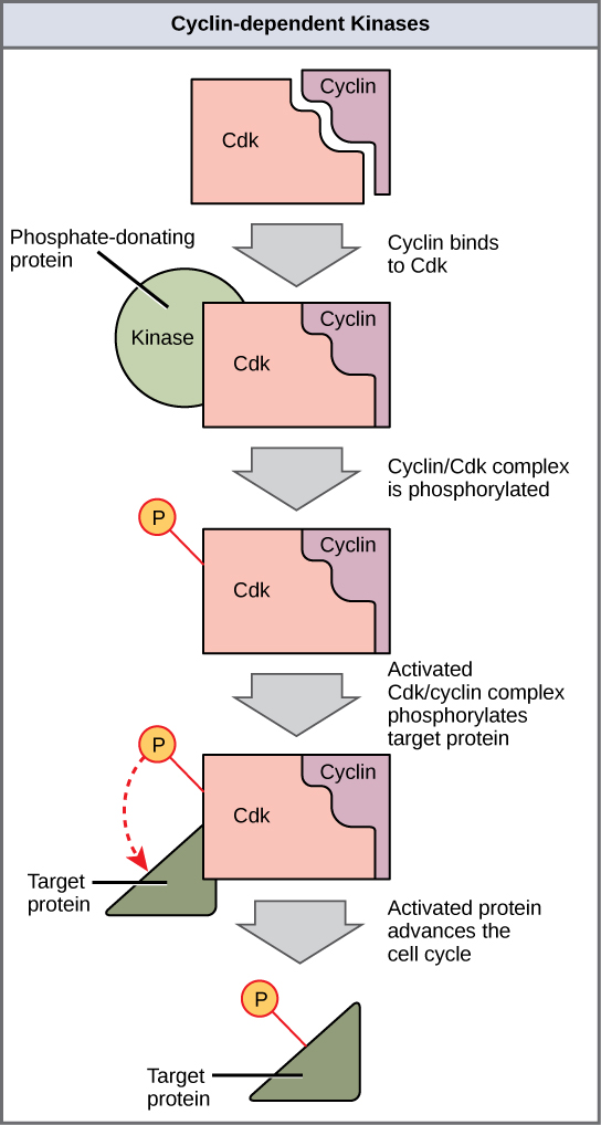
Two groups of proteins, called cyclins and cyclin-dependent kinases (CDKs), are termed positive regulators. They are responsible for the progress of the cell through the various checkpoints. The levels of the four cyclin proteins fluctuate throughout the cell cycle in a predictable pattern. Increases in the concentration of cyclin proteins are triggered by both external and internal signals. After the cell moves to the next stage of the cell cycle, the cyclins that were active in the previous stage are degraded by cytoplasmic enzymes.
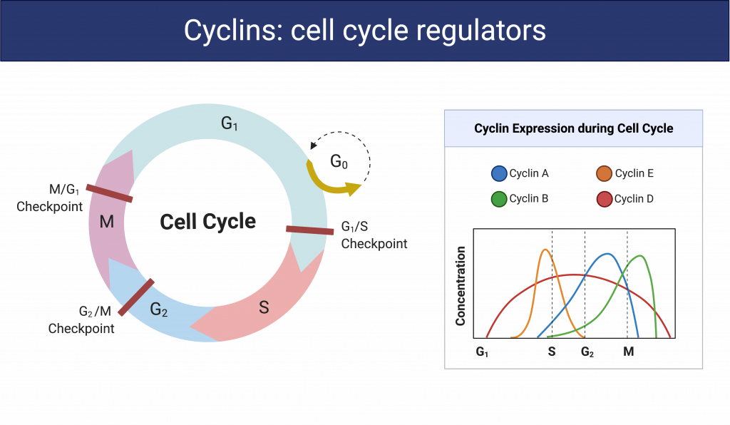
Cyclins regulate the cell cycle only when they are tightly bound to CDKs. To be fully active, the CDK/cyclin complex must also be phosphorylated in specific locations to activate the complex. Like all kinases, CDKs are enzymes (kinases) that in turn phosphorylate other proteins. Phosphorylation activates the protein by changing its shape. The proteins phosphorylated by CDKs are involved in advancing the cell to the next phase. The levels of CDK proteins are relatively stable throughout the cell cycle; however, the concentrations of cyclin fluctuate and determine when cyclin/CDK complexes form.
Because the cyclic fluctuations of cyclin levels are largely based on the timing of the cell cycle and not on specific events, regulation of the cell cycle usually occurs by either the CDK molecules alone or the cyclin/CDK complexes. Without a specific concentration of fully activated cyclin/CDK complexes, the cell cycle cannot proceed through the checkpoints.
Although the cyclins are the main regulatory molecules that determine the forward momentum of the cell cycle, there are several other mechanisms that fine-tune the progress of the cycle with negative, rather than positive, effects. These mechanisms essentially block the progression of the cell cycle until problematic conditions are resolved. Molecules that prevent the full activation of CDKs are called CDK inhibitors. Many of these inhibitor molecules directly or indirectly monitor a particular cell-cycle event. The block placed on CDKs by inhibitor molecules will not be removed until the specific event that the inhibitor monitors is completed.
Negative Regulation of the Cell Cycle
The second group of cell-cycle regulatory molecules are negative regulators, which stop the cell cycle. Remember that in positive regulation, active molecules cause the cycle to progress.
The best understood negative regulatory molecules are retinoblastoma protein (Rb), p53, and p21. Retinoblastoma proteins are a group of tumor suppressor proteins common in many cells. We should note here that the 53 and 21 designations refer to the functional molecular masses of the proteins (p) in kilodaltons. Much of what is known about cell-cycle regulation comes from research conducted with cells that have lost regulatory control. All three of these regulatory proteins were discovered to be damaged or non-functional in cells that had begun to replicate uncontrollably (i.e., became cancerous). In each case, the main cause of the unchecked progress through the cell cycle was a faulty copy of the regulatory protein.
Rb, p53, and p21 act primarily at the G1 checkpoint. p53 is a multi-functional protein that has a major impact on the commitment of a cell to division because it acts when there is damaged DNA in cells that are undergoing the preparatory processes during G1. If damaged DNA is detected, p53 halts the cell cycle and then recruits specific enzymes to repair the DNA. If the DNA cannot be repaired, p53 can trigger apoptosis, or cell suicide, to prevent the duplication of damaged chromosomes. As p53 levels rise, the production of p21 is triggered. p21 enforces the halt in the cycle dictated by p53 by binding to and inhibiting the activity of the cyclin/CDK complexes. As a cell is exposed to more stress, higher levels of p53 and p21 accumulate, making it less likely that the cell will move into the S phase.
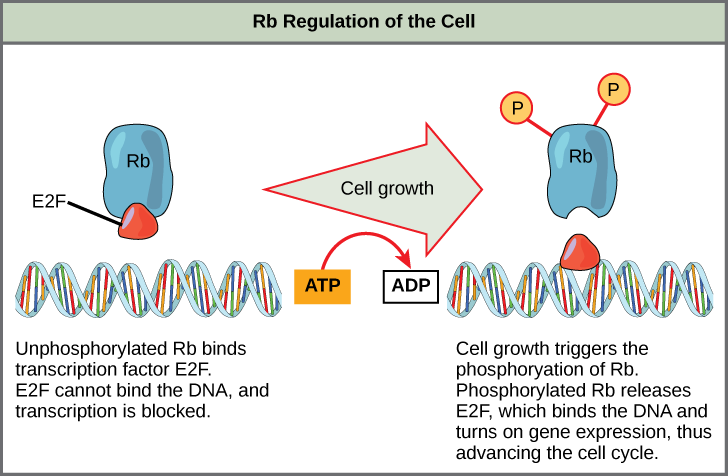
Rb, which largely monitors cell size, exerts its regulatory influence on other positive regulator proteins. In the active, dephosphorylated state, Rb binds to proteins called transcription factors, most commonly, E2F. Transcription factors “turn on” specific genes, allowing the production of proteins encoded by that gene. When Rb is bound to E2F, production of proteins necessary for the G1/S transition is blocked. As the cell increases in size, Rb is slowly phosphorylated until it becomes inactivated. Rb releases E2F, which can now turn on the gene that produces the transition protein, and this particular block is removed. For the cell to move past each of the checkpoints, all positive regulators must be “turned on,” and all negative regulators must be “turned off.”
one of a group of proteins that act in conjunction with cyclin-dependent kinases to help regulate the cell cycle by phosphorylating key proteins; the concentrations of cyclins fluctuate throughout the cell cycle
one of a group of protein kinases that helps to regulate the cell cycle when bound to cyclin; it functions to phosphorylate other proteins that are either activated or inactivated by phosphorylation
addition of a phosphate group
regulatory molecule that exhibits negative effects on the cell cycle by interacting with a transcription factor (E2F)
cell-cycle regulatory protein that regulates cell growth and monitors DNA damage; it halts the progression of the cell cycle in cases of DNA damage and may induce apoptosis
cell-cycle regulatory protein that inhibits the cell cycle; its levels are controlled by p53
protein that is a negative regulator of the cell cycle; prevents the cell from undergoing uncontrolled division
a protein that binds to DNA and regulates gene expression by either promoting or suppressing transcription
