
18.3 Meiosis
Overview
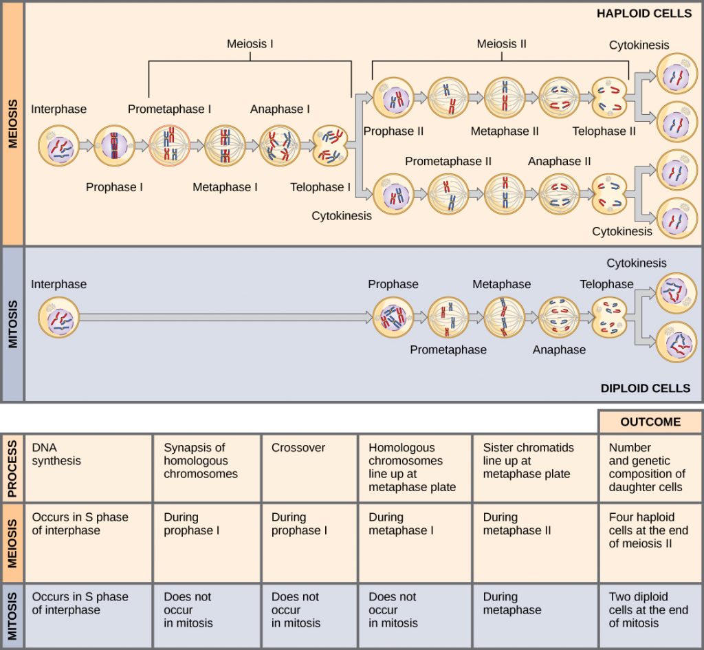
Meiosis is the nuclear division that forms haploid cells from diploid cells, and it employs many of the same cellular mechanisms as mitosis. However, as you have learned, mitosis produces daughter cells whose nuclei are genetically identical to the original parent nucleus. In mitosis, both the parent and the daughter nuclei are at the same “ploidy level”—diploid in the case of most multicellular animals. In meiosis, the daughter nuclei that result are half of the ploidy level of the parent cell. Most normally, the parent cell is diploid and the daughter cells are haploid.
To achieve this reduction in chromosome number, meiosis consists of one round of chromosome replication followed by two rounds of nuclear division. Because the events that occur during each of the division stages are analogous to the events of mitosis, the same stage names are assigned. However, because there are two rounds of division, the major process and the stages are designated with a “I” or a “II.” Thus, meiosis I is the first round of meiotic division and consists of prophase I, prometaphase I, and so on. Likewise, meiosis II (during which the second round of meiotic division takes place) includes prophase II, prometaphase II, and so on.
Meiosis and fertilization produce unique combinations of genetic material by three mechanisms — genetic recombination during prophase I, independent assortment during metaphase I, and random fertilization.
Meiosis I
Meiosis I separates homologous chromosomes into different daughter cells. The ploidy level of the daughter cells is half of the parent cell; for diploid organisms, the products of meiosis I are haploid.
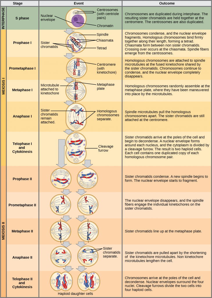
Interphase
Meiosis is preceded by an interphase consisting of G1, S, and G2 phases, which are nearly identical to the phases preceding mitosis. The G1 phase (the “first gap phase”) is focused on cell growth. During the S phase—the second phase of interphase—the cell copies or replicates the DNA of the chromosomes. Finally, in the G2 phase (the “second gap phase”) the cell undergoes the final preparations for meiosis.
During DNA duplication in the S phase, each chromosome is replicated to produce two identical sister chromatids that are held together at the centromere and are hence part of the same chromosome until anaphase II when they separate from one another.
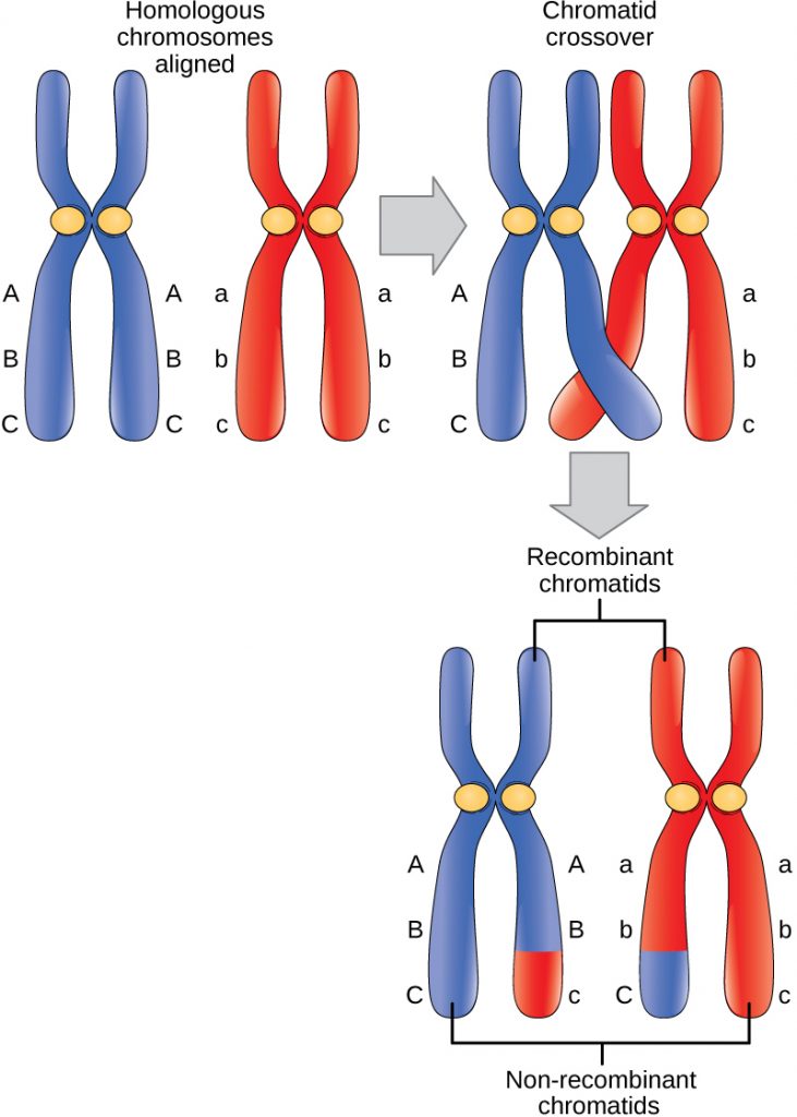
Prophase I and Crossing Over
Early in prophase I, before the chromosomes can be seen clearly with a microscope, the homologous chromosomes are attached at their tips to the nuclear envelope by proteins. As the nuclear envelope begins to break down, the proteins associated with homologous chromosomes bring the pair closer together. Recall that in mitosis, homologous chromosomes do not pair together. The tight pairing of the homologous chromosomes is called synapsis. In synapsis, the genes on the chromatids of the homologous chromosomes are aligned precisely with each other, which allows the exchange of chromosomal segments between homologous nonsister chromatids—a process called crossing over. Crossing over can be observed visually after the exchange as chiasmata (singular = chiasma).
The crossover events are the first source of genetic variation in the nuclei produced by meiosis. A single crossover event between homologous nonsister chromatids leads to a reciprocal exchange of equivalent DNA between a maternal chromosome and a paternal chromosome. When a recombinant sister chromatid is moved into a gamete cell it will carry some DNA from one parent and some DNA from the other parent. The recombinant chromatid has a combination of maternal and paternal genes that did not exist before the crossover. Crossover events can occur almost anywhere along the length of the synapsed chromosomes. Different cells undergoing meiosis will therefore produce different recombinant chromatids, with varying combinations of maternal and paternal genes. Multiple crossovers in an arm of the chromosome have the same effect, exchanging segments of DNA to produce genetically recombined chromosomes.
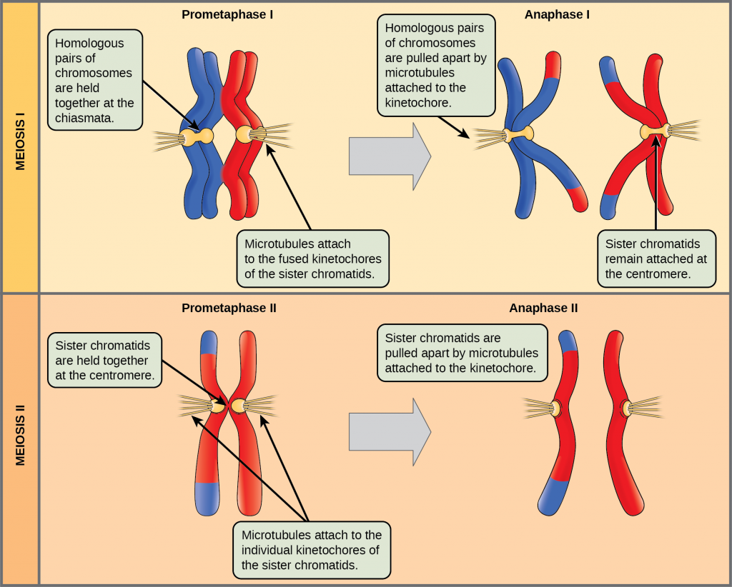
Prometaphase I
The key event in prometaphase I is the attachment of the spindle fiber microtubules to the kinetochore proteins at the centromeres. The microtubules from each pole move toward the middle of the cell and attach to one of the kinetochores of the two fused homologous chromosomes. Each member of the homologous pair attaches to a microtubule extending from opposite poles of the cell so that in the next phase, the microtubules can pull the homologous pair apart.
Metaphase I
During metaphase I, the pairs of homologous chromosomes are arranged at the metaphase plate—roughly in the midline of the cell, with the kinetochores facing opposite poles. The homologous pairs orient themselves randomly at the equator. For example, if the two homologous members of chromosome 1 are labeled a and b, then the chromosomes could line up a-b or b-a. This is important in determining the genes carried by a gamete, as each will only receive one chromosome of each homologous chromosome pair. (Recall that after crossing over takes place, homologous chromosomes are not identical. They contain slight differences in their genetic information, causing each gamete to have a unique genetic makeup.)
The randomness in the alignment of recombined chromosomes at the metaphase plate, coupled with the crossing over events between nonsister chromatids, are responsible for much of the genetic variation in the offspring. To clarify this further, remember that the homologous chromosomes of a sexually reproducing organism are originally inherited as two separate sets, one from each parent. Using humans as an example, one set of 23 chromosomes is present in the egg donated by the mother. The father provides the other set of 23 chromosomes in the sperm that fertilizes the egg. Every cell of the multicellular offspring has copies of the original two sets of homologous chromosomes. In prophase I of meiosis, the homologous chromosomes form the tetrads. In metaphase I, these pairs line up at the midway point between the two poles of the cell to form the metaphase plate. Because there is an equal chance that a microtubule fiber will encounter a maternally or paternally inherited chromosome, the arrangement of the tetrads at the metaphase plate is random. Thus, any maternally inherited chromosome may face either pole. Likewise, any paternally inherited chromosome may also face either pole. The orientation of each tetrad is independent of the orientation of the other 22 tetrads.
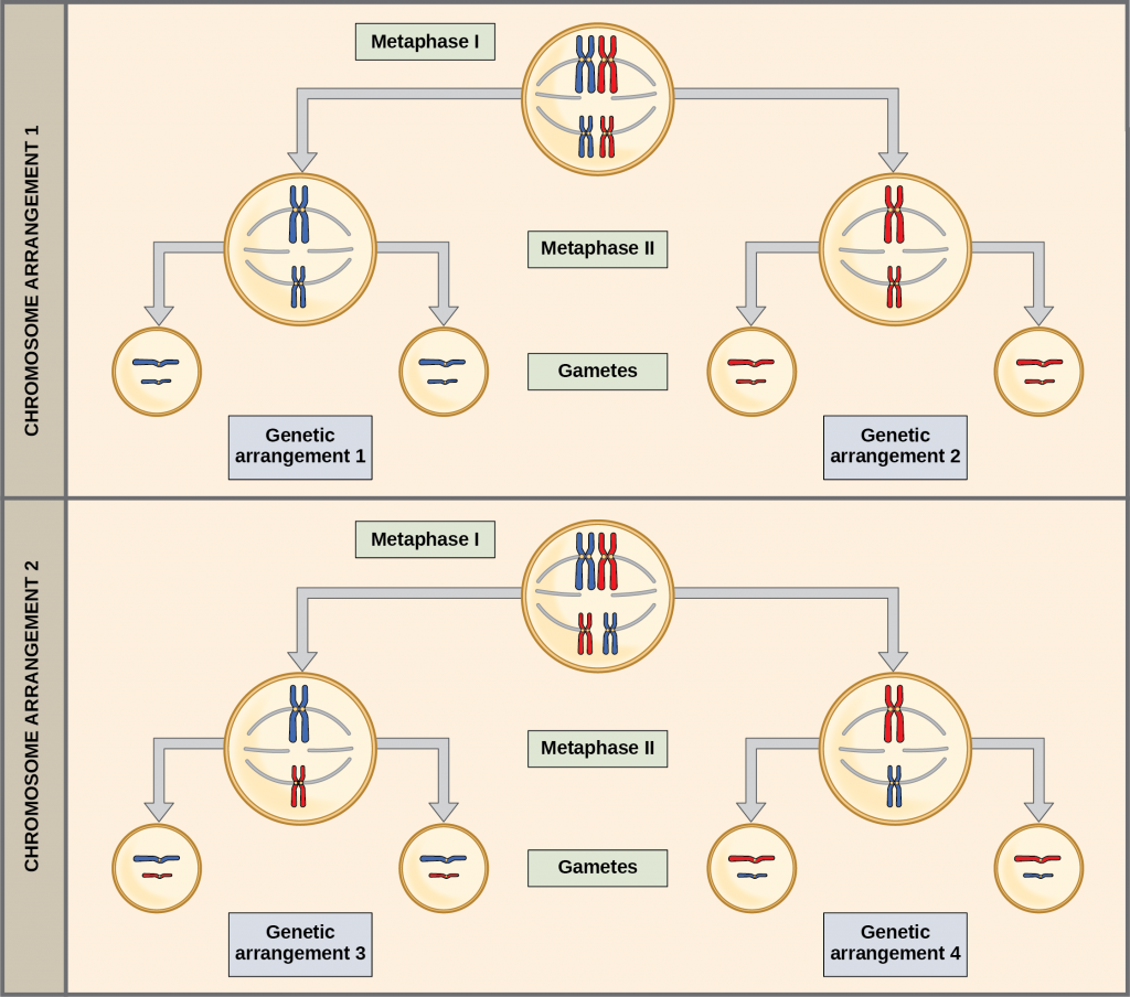
This event—the independent assortment of homologous chromosomes at the metaphase plate—is the second mechanism that introduces variation into the gametes or spores. In each cell that undergoes meiosis, the arrangement of the tetrads is different. The number of variations is dependent on the number of chromosomes making up a set. There are two possibilities for orientation at the metaphase plate; the possible number of alignments therefore equals 2n in a diploid cell, where n is the number of chromosomes per haploid set. Humans have 23 chromosome pairs, which results in over eight million (223) possible genetically-distinct gametes just from the random alignment of chromosomes at the metaphase plate. This number does not include the variability that was previously described (produced by crossing over between the nonsister chromatids). Given these two mechanisms, it is nearly impossible that any two haploid cells resulting from meiosis will have the same genetic composition.
Anaphase I
In anaphase I, the microtubules pull the linked chromosomes apart. The sister chromatids remain tightly bound together at the centromere. The chiasmata are broken in anaphase I as the microtubules attached to the fused kinetochores pull the homologous chromosomes apart.
Telophase I and Cytokinesis
In telophase, the separated chromosomes arrive at opposite poles. The remainder of the typical telophase events may or may not occur, depending on the species. In some organisms, the chromosomes “decondense” and nuclear envelopes form around the separated sets of chromatids produced during telophase I. In other organisms, cytokinesis—the physical separation of the cytoplasmic components into two daughter cells—occurs without reformation of the nuclei.
Two haploid cells are the result of the first meiotic division of a diploid cell. The cells are haploid because at each pole, there is just one of each pair of the homologous chromosomes. Therefore, only one full set of the chromosomes is present. This is why the cells are considered haploid—there is only one chromosome set, even though each chromosome still consists of two sister chromatids. Recall that sister chromatids are merely duplicates of one of the two homologous chromosomes (except for changes that occurred during crossing over). In meiosis II, these two sister chromatids will separate, creating four haploid daughter cells.
Meiosis II
In some species, cells enter a brief interphase, or interkinesis, before entering meiosis II. Interkinesis lacks an S phase, so chromosomes are not duplicated. During meiosis II, the sister chromatids within the two daughter cells separate, forming four new haploid gametes. The mechanics of meiosis II are similar to mitosis, except that each dividing cell has only one set of homologous chromosomes, each with two chromatids. Therefore, each cell has half the number of sister chromatids to separate out as a diploid cell undergoing mitosis. In terms of chromosomal content, cells at the start of meiosis II are similar to haploid cells in G2, preparing to undergo mitosis.
The stages of meiosis II — prophase II, prometaphase II, metaphase II, anaphase II, and telophase II — are similar to the equivalent stages of mitosis.
Comparing Meiosis and Mitosis
Mitosis and meiosis are both forms of division of the nucleus in eukaryotic cells. They share some similarities, but also exhibit a number of important and distinct differences that lead to very different outcomes. Mitosis is a single nuclear division that results in two nuclei that are usually partitioned into two new cells. The nuclei resulting from a mitotic division are genetically identical to the original nucleus. They have the same number of sets of chromosomes: one set in the case of haploid cells and two sets in the case of diploid cells. In contrast, meiosis consists of two nuclear divisions resulting in four nuclei that are usually partitioned into four new, genetically distinct cells. The four nuclei produced during meiosis are not genetically identical, and they contain one chromosome set only. This is half the number of chromosome sets in the original cell, which was diploid.
(also, karyokinesis) period of the cell cycle during which the duplicated chromosomes are separated into identical nuclei; includes prophase, prometaphase, metaphase, anaphase, and telophase
first round of meiotic cell division; referred to as reduction division because the daughter cells have half the number of chromosomes as the parent cell
second round of meiotic cell division following meiosis I; sister chromatids are separated into individual chromosomes, and the result is (usually) four unique haploid cells
exchange of genetic material between chromosomes of two different individuals
individual alleles or chromosomes assort into gametes independently of other alleles or chromosomes
the concept that gametes unite independent of their genetic makeup; for example, in humans it is random which sperm fertilizes which egg
formation of a close association between homologous chromosomes during prophase I
exchange of genetic material between homologous chromosomes resulting in chromosomes that incorporate genes from both parents of the organism
(singular, chiasma) the structure that forms at the crossover points after genetic material is exchanged
division of the cytoplasm following mitosis that forms two daughter cells
(also, interphase II) brief period of rest between meiosis I and meiosis II
