
13 Chapter 13: Nervous System
Chapter Outline
- 13.1 Neurons and Glial Cells
- 13.2 How Neurons Communicate
- 13.3 The Central Nervous System
- 13.4 The Peripheral Nervous System

Introduction to the Nervous System
13.1 Neurons and Glial Cells
The nervous system is made up of neurons, specialized cells that can receive and transmit chemical or electrical signals, and glia, cells that provide support functions for the neurons by playing an information processing role that is complementary to neurons. A neuron can be compared to an electrical wire-it transmits a signal from one place to another. Glia can be compared to the workers at the electric company who make sure wires go to the right places, maintain the wires, and take down wires that are broken. Although glia have been compared to workers, recent evidence suggests that also take on some of the signaling functions of neurons.
13.1.1 Neurons
The nervous system of the common laboratory fly, Drosophila melanogaster, contains around 100,000 neurons, the same number as a lobster. This number compares to 75 million in the mouse and 300 million in the octopus. A human brain contains around 86 billion neurons. Despite these very different numbers, the nervous systems of these animals control many of the same behaviors-from basic reflexes to more complicated behaviors like finding food and courting mates. The ability of neurons to communicate with each other as well as with other types of cells underlies all of these behaviors.
Most neurons share the same cellular components. But neurons are also highly specialized-different types of neurons have different sizes and shapes that relate to their functional roles.
13.1.2 Parts of a Neuron
Like other cells, each neuron has a cell body that contains a nucleus, mitochondria, and other cellular components. Neurons also contain unique structures, illustrated in Figure 13.2 for receiving and sending the electrical signals that make neuronal communication possible. Dendrites are tree-like structures that extend away from the cell body to receive messages from other neurons at specialized junctions called synapses.
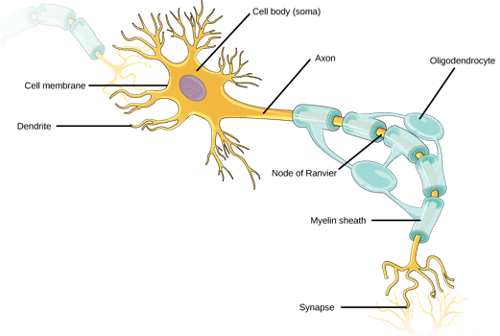
Once a signal is received by the dendrite, it then travels passively to the cell body. The cell body contains a specialized structure, the axon hillock that integrates signals from multiple synapses and serves as a junction between the cell body and an axon. An axon is a tube-like structure that propagates (carries) the signal to specialized endings called axon terminals. These terminals in turn synapse on other neurons, muscle, or target organs. Chemicals released at axon terminals allow signals to be communicated to these other cells. Some axons are covered with myelin, which acts as an insulator to minimize loss of the electrical signal as it travels down the axon; minimizing this loss of signal greatly increasing the speed on conduction. This insulation is important as the axon from a human motor neuron can be as long as a meter, such as the neurons that travel from the base of the spine to the toes. The myelin sheath is not actually part of the neuron. Myelin is produced by glial cells.
It is important to note that a single neuron does not act alone-neuronal communication depends on the connections that neurons make with one another (as well as with other cells, like muscle cells). Dendrites from a single neuron may receive signals from many other neurons. For example, dendrites from a Purkinje cell in the cerebellum are thought to receive contact from as many as 100,000 other neurons.
13.1.3 Glia
While glia are often thought of as the supporting cast of the nervous system, the number of glial cells in the brain actually outnumbers the number of neurons by a factor of ten. Neurons would be unable to function without the vital roles that are fulfilled by these glial cells. Glia guide developing neurons to their destinations, buffer ions and chemicals that would otherwise harm neurons, and provide myelin sheaths around axons. Scientists have recently discovered that they also play a role in responding to nerve activity and modulating communication between nerve cells. When glia do not function properly, the result can be disastrous-most brain tumors are caused by mutations in glia.
13.1.4 Section Summary
The nervous system is made up of neurons and glia. Neurons are specialized cells that are capable of sending electrical as well as chemical signals. Most neurons contain dendrites, which receive these signals, and axons that send signals to other neurons or tissues. There are four main types of neurons: unipolar, bipolar, multipolar, and pseudounipolar neurons. Glia are non-neuronal cells in the nervous system that support neuronal development and signaling. There are several types of glia that serve different functions.
13.2 How Neurons Communicate
All functions performed by the nervous system-from a simple motor reflex to more advanced functions like making a memory or a decision-require neurons to communicate with one another. While humans use words and body language to communicate, neurons use electrical and chemical signals. Just like a person in a committee, one neuron usually receives and synthesizes messages from multiple other neurons before “making the decision” to send the message on to other neurons.
13.2.1 Nerve Impulse Transmission within a Neuron
For the nervous system to function, neurons must be able to send and receive signals. A neuron can receive input from other neurons and, if this input is strong enough, send the signal to downstream neurons. The signal is either excitatory (stimulating the neuron to send another impulse) or inhibitory (suppressing or preventing the neuron from sending a signal). Transmission of a signal between neurons is generally carried by a chemical called a neurotransmitter. Transmission of a signal within a neuron, from the dendrite (receiver of signal) to the axon terminal (sender of the subsequent signal) is carried by an electrical impulse called an action potential.
Action Potential
An action potential is an electrical impulse, specifically a reversal in charge from negative to positive. These charges are possible because each neuron’s plasma membrane has a voltage difference (either positive or negative) between the inside and the outside of the cell). The charge of this membrane can change in response to neurotransmitter molecules released from other neurons. The charge can also change as the result of environmental stimuli.
When a neuron is not receiving a signal, it has a negative charge called the resting membrane potential. When neurotransmitter molecules bind to receptors located on a neuron’s dendrites, ion channels open (ions are atoms that have either a positive or negative charge). At excitatory synapses, this opening allows positive ions to enter the neuron and results in depolarization (reversal in charge, in this case from negative to positive) of the plasma membrane (Figure 13.3).
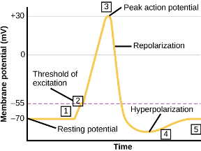
This electrical impulse from negative to positive propagates (travels) down the axon of the neuron, carrying the signal to the next target such as another neuron or a muscle (Figure 13.4). Then, the cell “resets” itself back to the negative resting membrane potential – this process is called repolarization.
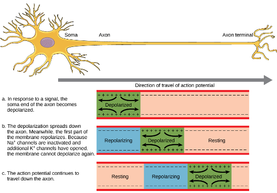
13.2.2 Myelin and the Propagation of the Action Potential
For an action potential to communicate information to another neuron, it must travel along the axon and reach the axon terminals where it can initiate neurotransmitter release. The speed of conduction of an action potential along an axon is influenced by both the diameter of the axon and the axon’s resistance to ion leakage. Myelin acts as an insulator that prevents current from leaving the axon, much like a the plastic wrapping on the electrical cords of computers and appliances. This insulation increases the speed of action potential conduction. In demyelinating diseases like multiple sclerosis, action potential conduction slows because ions leak from previously insulated axon areas. If the demyelination from multiple sclerosis is severe enough, the action potential is unable to propagate down the axon, which results in paralysis of the muscle that the neuron is attempting to signal.
13.2.3 Synaptic Transmission
Neurons are not physically in contact with each other. There is a synapse or “gap” between them and this is the place where information is transmitted from one neuron to another. Synapses usually form between axon terminals and dendritic spines. The neuron transmitting the signal is called the presynaptic neuron, and the neuron receiving the signal is called the postsynaptic neuron. There are two types of synapses: chemical and electrical.
Chemical Synapse
When an action potential reaches the axon terminal it depolarizes the membrane and opens voltage-gated Na+ channels. Na+ ions enter the cell, further depolarizing the presynaptic membrane. This depolarization causes voltage-gated Ca + channels to open. Calcium ions entering the cell initiate a signaling cascade that causes small membrane-bound vesicles, called synaptic vesicles, containing neurotransmitter molecules to fuse with the presynaptic membrane. Synaptic vesicles are shown in Figure 13.5, which is an image from a scanning electron microscope.

Fusion of a vesicle with the presynaptic membrane causes neurotransmitter to be released into the synaptic cleft, the extracellular space between the presynaptic and postsynaptic membranes, as illustrated in Figure 13.6 . The neurotransmitter diffuses across the synaptic cleft and binds to receptor proteins on the postsynaptic membrane.
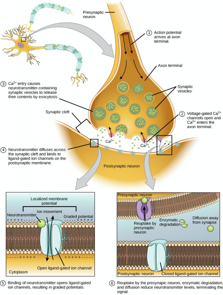
The binding of a specific neurotransmitter causes particular ion channels, in this case chemically-gated channels, on the postsynaptic membrane to open. Neurotransmitters can either have excitatory or inhibitory effects on the postsynaptic membrane. For example, when the neurotransmitter acetylcholine is released at the synapse between a nerve and muscle (called the neuromuscular junction) by a presynaptic neuron, it causes postsynaptic Na+ channels to open. Na+ enters the postsynaptic cell and causes the postsynaptic membrane to depolarize. This depolarization makes the postsynaptic neuron more likely to fire an action potential. Release of neurotransmitter at inhibitory synapses causes a hyperpolarization (the charge becomes more negative) of the presynaptic membrane. For example, when the neurotransmitter GABA (gamma-aminobutyric acid) is released from a presynaptic neuron, it binds to and opens Cl- channels. Cl- ions enter the cell and hyperpolarizes the membrane, making the neuron less likely to fire an action potential.
Once neurotransmission has occurred, the neurotransmitter must be removed from the synaptic cleft so the postsynaptic membrane can “reset” and be ready to receive another signal. This can be accomplished in three ways: the neurotransmitter can diffuse away from the synaptic cleft, it can be degraded by enzymes in the synaptic cleft, or it can be recycled (sometimes called reuptake) by the presynaptic neuron. Several drugs act at this step of neurotransmission. For example, some drugs that are given to Alzheimer’s patients work by inhibiting acetylcholinesterase, the enzyme that degrades acetylcholine. This inhibition of the enzyme essentially increases neurotransmission at synapses that release acetylcholine. Once released, the acetylcholine stays in the cleft and can continually bind and unbind to postsynaptic receptors.
13.2.4 Section Summary
Neurons have charged membranes because there are different concentrations of ions inside and outside of the cell. Voltage-gated ion channels control the movement of ions into and out of a neuron. When a neuronal membrane is depolarized to at least the threshold of excitation, an action potential is fired. The action potential is then propagated along an axon to the axon terminals. In a chemical synapse, the action poten- tial causes release of neurotransmitter molecules into the synaptic cleft. Through binding to postsynaptic receptors, the neurotransmitter can cause excitatory or inhibitory postsynaptic potentials by depolarizing or hyperpolarizing, respectively, the postsynaptic membrane.
13.3 The Central Nervous System
The central nervous system (CNS) is made up of two organs: the brain and the spinal cord. These organs receive information from the outside world (sensory), process that information, and send a response to the body (motor). The peripheral nervous system (PNS) is made of all the nerves that arise from the brain and spinal cord, and then travel throughout the body. The PNS carries both sensory information from the body to the brain and spinal cord, as well as motor information from the brain and spinal cord to the body (motor).
13.3.1 Brain
The brain is the part of the central nervous system that is contained in the cranial cavity of the skull. It includes the cerebral cortex, limbic system, thalamus, hypothalamus, cerebellum, brainstem, and retinas.
Cerebral Cortex
The outermost and largest part of the brain is a thick piece of nervous system tissue called the cerebral cortex. It is divided into the left and the right hemispheres. A thick fiber bundle called the corpus callosum (corpus = “body”; callosum = “tough”) connects the two hemispheres. Many of the functions of the body are processed in the opposite hemisphere – the right side of the body is controlled by motor areas in the left hemisphere. Likewise, sensory information such as images from the left visual field of the eyes are processed in the right hemisphere. Although there are some brain functions that are specialized to only one hemisphere, the functions of the two hemispheres are largely redundant. In fact, sometimes (very rarely) an entire hemisphere is removed to treat severe epilepsy. While patients do suffer some deficits following the surgery, they can have surprisingly few problems, especially when the surgery is performed on children who have very immature nervous systems. In other surgeries to treat severe epilepsy, the corpus callosum is cut instead of removing an entire hemisphere. This causes a condition called split-brain, which gives insights into unique functions of the two hemispheres. For example, when an object is presented to patients’ left visual field, they may be unable to verbally name the object (and may claim to not have seen an object at all). This is because the visual input from the left visual field crosses and enters the right hemisphere and cannot then signal to the speech center, which generally is found in the left side of the brain. Remarkably, if a split-brain patient is asked to pick a specific object out of a group of objects with the left hand, the patient will be able to do so but will still be unable to verbally identify it.
Each hemisphere contains regions called lobes that are involved in different functions. Each hemisphere of the mammalian cerebral cortex can be broken down into four functionally and spatially defined lobes: frontal, parietal, temporal, and occipital (Figure 13.7).
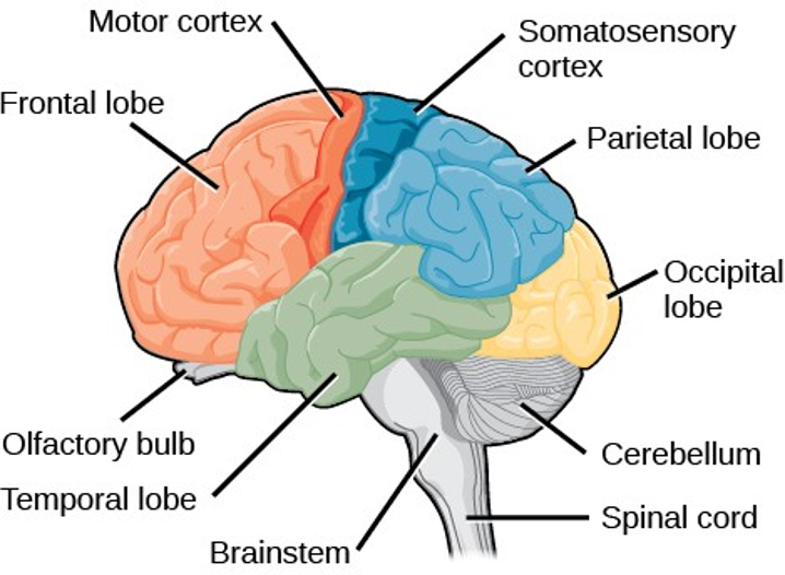
The frontal lobe is located at the front of the brain, over the eyes. This lobe contains the olfactory bulb, which processes smells. The frontal lobe also contains the motor cortex, which is important for planning and implementing movement. Areas within the motor cortex map to different muscle groups. Neurons in the frontal lobe also control cognitive functions like maintaining attention, speech, and decision-making. Studies of humans who have damaged their frontal lobes show that parts of this area are involved in personality, socialization, and assessing risk. The parietal lobe is located at the top of the brain. Neurons in the parietal lobe are involved in speech and also reading. Two of the parietal lobe’s main functions are processing somatosensation-touch sensations like pressure, pain, heat, cold-and processing proprioception-the sense of how parts of the body are oriented in space. The parietal lobe contains a somatosensory map of the body similar to the motor cortex. The occipital lobe is located at the back of the brain. It is primarily involved in vision-seeing, recognizing, and identifying the visual world. The temporal lobe is located at the base of the brain and is primarily involved in processing and interpreting sounds. It also contains the hippocampus (named from the Greek for “seahorse,” which it resembles in shape) a structure that processes memory formation. The role of the hippocampus in memory was partially determined by studying one famous epileptic patient, HM, who had both sides of his hippocampus removed in an attempt to cure his epilepsy. His seizures went away, but he could no longer form new memories (although he could remember some facts from before his surgery and could learn new motor tasks).
Other Structures of the Brain
The thalamus is directly underneath the cerebral cortex and acts as a gateway to and from the cortex. Motor impulses from the cortex travel down through it to the spinal cord and then to the peripheral nervous system, which sends the signal to muscles and glands in the body. Sensory impulses travel in the opposite direction carrying information from the body to the brain. It also receives feedback from the cortex. This feedback mechanism can modulate conscious awareness of sensory and motor inputs depending on the attention and arousal state of the animal. The thalamus helps regulate consciousness, arousal, and sleep states.
Below the thalamus is the hypothalamus. The hypothalamus controls the endocrine system by sending signals to the pituitary gland. Among other functions, the hypothalamus is the body’s thermostat-it makes sure the body temperature is kept at appropriate levels. Neurons within the hypothalamus also regulate circadian rhythms, sometimes called sleep cycles.
The limbic system is a connected set of structures that regulates emotion, as well as behaviors related to fear and motivation. It plays a role in memory formation and includes parts of the thalamus and hypothalamus as well as the hippocampus. One important structure within the limbic system is a temporal lobe structure called the amygdala. The two amygdala (one on each side) are important both for the sensation of fear and for recognizing fearful faces.
The cerebellum (cerebellum = “little brain”) sits at the base of the brain on top of the brainstem. The cerebellum controls balance and aids in coordinating movement and learning new motor tasks. The cerebellum of birds is large compared to other vertebrates because of the coordination required by flight.
The brainstem connects the rest of the brain with the spinal cord and regulates some of the most important and basic functions of the nervous system including breathing, swallowing, digestion, sleeping, walking, and sensory and motor information integration.
13.3.1 Spinal cord
Connecting to the brainstem and extending down the body through the spinal column is the spinal cord. The spinal cord is a thick bundle of nerve tissue that carries information about the body to the brain and from the brain to the body. The spinal cord is contained within the meninges and the bones of the vertebral column but is able to communicate signals to and from the body through its connections with spinal nerves (part of the peripheral nervous system). A cross-section of the spinal cord looks like a white oval containing a gray butterfly-shape (Figure 17.16). Myelinated axons make up the “white matter” and neuron and glia cell bodies (and interneurons) make up the “gray matter.” Axons and cell bodies in the dorsal spinal cord convey mostly sensory information from the body to the brain. Axons and cell bodies in the ventral spinal cord primarily transmit signals controlling movement from the brain to the body.
The spinal cord also controls motor reflexes. These reflexes are quick, unconscious movements-like automatically removing a hand from a hot object. Reflexes are so fast because they involve local synaptic connections. For example, the knee reflex that a doctor tests during a routine physical is controlled by a single synapse between a sensory neuron and a motor neuron. While a reflex may only require the involvement of one or two synapses, synapses with interneurons in the spinal column transmit information to the brain to convey what happened (the knee jerked, or the hand was hot).
13.4 The Peripheral Nervous System
The peripheral nervous system (PNS) is the connection between the central nervous system and the rest of the body. The PNS can be broken down into the autonomic nervous system, which controls bodily functions without conscious control, and the sensory-somatic nervous system, which transmits sensory information from the skin, muscles, and sensory organs to the CNS and sends motor commands from the CNS to the muscles.
Autonomic Nervous System
The autonomic nervous system serves as the relay between the CNS and the internal organs. It controls the lungs, the heart, smooth muscle, and exocrine and endocrine glands and vicera of the abdominal cavity. The autonomic nervous system controls these organs largely without conscious control; it can continuously monitor the conditions of these different systems and implement changes as needed. Signaling to the target tissue usually involves two synapses: a preganglionic neuron (originating in the CNS) synapses to a neuron in a ganglion that, in turn, synapses on the target organ (Figure 17.17). There are two divisions of the autonomic nervous system that often have opposing effects: the sympathetic nervous system and the parasympathetic nervous system.
The sympathetic nervous system is responsible for the immediate responses an animal makes when it encounters a dangerous situation. One way to remember this is to think of the “fight-or-flight” response a person feels when encountering a snake (“snake” and “sympathetic” both begin with “s”). Examples of functions controlled by the sympathetic nervous system include an accelerated heart rate and inhibited digestion. These functions help prepare an organism’s body for the physical strain required to escape a potentially dangerous situation or to fend off a predator.
While the sympathetic nervous system is activated in stressful situations, the parasympathetic nervous system allows an animal to “rest and digest.” One way to remember this is to think that during a restful situation like a picnic, the parasympathetic nervous system is in control (“picnic” and “parasympathetic” both start with “p”). Parasympathetic preganglionic neurons have cell bodies located in the brainstem and in the sacral (toward the bottom) spinal cord (Figure 17.18). The parasympathetic nervous system resets organ function after the sympathetic nervous system is activated including slowing of heart rate, lowered blood pressure, and stimulation of digestion.
The sensory-somatic nervous system is made up of cranial and spinal nerves and contains both sensory and motor neurons. Sensory neurons transmit sensory information from the skin, skeletal muscle, and sensory organs to the CNS. Motor neurons transmit messages about desired movement from the CNS to the muscles to make them contract. Without its sensory-somatic nervous system, an animal would be unable to process any information about its environment (what it sees, feels, hears, and so on) and could not control motor movements. Unlike the autonomic nervous system, which usually has two synapses between the CNS and the target organ, sensory and motor neurons usually have only one synapse-one ending of the neuron is at the organ and the other directly contacts a CNS neuron.
Chapter Review Questions
Exercise 1
Neurons contain_____________ , which can receive signals from other neurons.
- axons
- mitochondria
- dendrites
- Golgi bodies
Exercise 2
A(n)_____________ neuron has one axon and multiple dendrites.
- unipolar
- bipolar
- multipolar
- pseudounipolar
Exercise 3
How are neurons similar to other cells? How are they unique?
Exercise 4
Multiple sclerosis causes demyelination of axons in the brain and spinal cord. Why is this prob- lematic?
Exercise 5
The part of the brain that is responsible for coordination during movement is the________ _.
- limbic system
- thalamus
- cerebellum
- parietal lobe
Exercise 6
Which part of the nervous system directly controls the digestive system?
- parasympathetic nervous system
- central nervous system
- spinal cord
- sensory-somatic nervous system
Free Response
Exercise 17.4.3
What are the main functions of the spinal cord?
Exercise 17.4.4
What are the main differences between the sympathetic and parasympathetic branches of the autonomic nervous system?
Exercise 17.4.5
What are the main functions of the sensory-somatic nervous system?
Exercise 17.3.2
For a neuron to fire an action potential, its membrane must reach___________ _.
- hyperpolarization
- the threshold of excitation
- the refractory period
- inhibitory postsynaptic potential
Exercise 17.3.3
After an action potential, the opening of additional voltage-gated_____________ channels and the
inactivation of sodium channels, cause the membrane to return to its resting membrane potential.
- sodium
- potassium
- calcium
- chloride
17.1.1 Free Response
Exercise 17.3.4
How does myelin aid propagation of an action potential along an axon? How do the nodes of Ranvier help this process?
Exercise 17.3.5
What are the main steps in chemical neurotransmission?
Solutions to Exercises
to Exercise 17.2.1 (p. 357) C
to Exercise 17.2.2 (p. 357) C
to Exercise 17.2.3 (p. 357)
Neurons contain organelles common to all cells, such as a nucleus and mitochondria. They are unique because they contain dendrites, which can receive signals from other neurons, and axons that can send these signals to other cells.
to Exercise 17.2.4 (p. 357)
Myelin provides insulation for signals traveling along axons. Without myelin, signal transmission can slow down and degrade over time. This would slow down neuronal communication across the nervous system and affect all downstream functions.
to Exercise 17.3.1 (p. 367)
Figure 17.8 Potassium channel blockers slow the repolarization phase, but have no effect on depolarization. to Exercise 17.3.2 (p. 367)
B
to Exercise 17.3.3 (p. 368) B
to Exercise 17.3.4 (p. 368)
Myelin prevents the leak of current from the axon. Nodes of Ranvier allow the action potential to be regenerated at specific points along the axon. They also save energy for the cell since voltage-gated ion channels and sodium-potassium transporters are not needed along myelinated portions of the axon.
to Exercise 17.3.5 (p. 368)
An action potential travels along an axon until it depolarizes the membrane at an axon terminal. Depo- larization of the membrane causes voltage-gated Ca + channels to open and Ca + to enter the cell. The intracellular calcium influx causes synaptic vesicles containing neurotransmitter to fuse with the presynaptic membrane. The neurotransmitter diffuses across the synaptic cleft and binds to receptors on the postsynap- tic membrane. Depending on the specific neurotransmitter and postsynaptic receptor, this action can cause positive (excitatory postsynaptic potential) or negative (inhibitory postsynaptic potential) ions to enter the cell.
to Exercise 17.4.1 (p. 376) C
to Exercise 17.4.2 (p. 376) A
to Exercise 17.4.3 (p. 376)
The spinal cord transmits sensory information from the body to the brain and motor commands from the brain to the body through its connections with peripheral nerves. It also controls motor reflexes.
to Exercise 17.4.4 (p. 376)
The sympathetic nervous system prepares the body for “fight or flight,” whereas the parasympathetic nervous system allows the body to “rest and digest.” Sympathetic neurons release norepinephrine onto target organs; parasympathetic neurons release acetylcholine. Sympathetic neuron cell bodies are located in sympathetic ganglia. Parasympathetic neuron cell bodies are located in the brainstem and sacral spinal cord. Activation of the sympathetic nervous system increases heart rate and blood pressure and decreases digestion and blood flow to the skin. Activation of the parasympathetic nervous system decreases heart rate and blood pressure and increases digestion and blood flow to the skin.
to Exercise 17.4.5 (p. 377)
The sensory-somatic nervous system transmits sensory information from the skin, muscles, and sensory organs to the CNS. It also sends motor commands from the CNS to the muscles, causing them to contract.
Media Attributions
- Figure 12.1
- Figure 12.2
- Figure 12.3
- Figure 12.4
- Figure 12.5
- Figure 12.6
- Figure 12.7
