
20 Chapter 20: Muscular System
Learning Objectives
After studying this chapter you should be able to:
- Understand the structure of muscle tissue by:
- a. Recognizing that muscles contract (shorten) to generate force.
- b. Distinguishing among the basic properties of the three types of muscle tissue.
- Name the functions (specific actions) and body regions of the major muscles of the body as indicated in the table below, and match the name with the muscle.
20.1 Muscle Types and Structure
Muscle cells are specialized for contraction. Muscles allow for motions such as walking, and they also facilitate bodily processes such as respiration and digestion. The body contains three types of muscle tissue: skeletal muscle, cardiac muscle, and smooth muscle (Figure 20.1).

Skeletal muscles, which attach to bones or skin, control locomotion and any movement that can be voluntarily controlled. Skeletal muscles are long and cylindrical in appearance; when viewed under a microscope, skeletal muscle tissue has a striped or striated appearance. The striations are caused by the regular arrangement of contractile proteins (actin and myosin). Actin is a filamentous contractile protein that interacts with another filamentous protein called myosin for muscle contraction to occur. Skeletal muscle also has multiple nuclei present in a single cell.
Smooth muscle is found in the walls of hollow organs such as the intestines, stomach, and urinary bladder, and around passages such as the airways and blood vessels. Smooth muscle has no striations, is not under voluntary control (it’s under the control of the autonomic nervous system), has only one nucleus per cell, is tapered at both ends, and is called involuntary muscle.
Cardiac muscle is only found in the heart, and cardiac muscle contractions pump blood throughout the body and maintain blood pressure. Like skeletal muscle, cardiac muscle is striated, but unlike skeletal muscle, cardiac muscle cannot be consciously controlled and is called involuntary muscle (it’s under the control of the autonomic nervous system like smooth muscle). Cardiac muscle cells can have more than one nucleus per cell and are branched.
Skeletal Muscle Fiber Structure
Each skeletal muscle fiber is a skeletal muscle cell. These cells are incredibly large, with diameters of up to 100 µm and lengths of up to 30 cm. Within each muscle fiber are myofibrils—long cylindrical structures that lie parallel to the muscle fiber. Myofibrils run the entire length of the muscle fiber, and because they are only approximately 1.2 µm in diameter, hundreds to thousands can be found inside one muscle fiber. When the myofibrils shorten, the entire muscle cell contracts and generates force, pulling on the tendons that attach the muscle to the bone and moving the bone. (Figure 20.2).

Myofibrils are composed of smaller structures called myofilaments. There are two main types of filaments: thick filaments and thin filaments; each has different compositions and locations. Thick filaments (composed of the protein myosin) and thin filaments (composed of the protein actin) (see Figure 20.3).

20.2 Muscle Actions
Synovial joints allow the body a tremendous range of movements. Each movement at a synovial joint results from the contraction or relaxation of the muscles that are attached to the bones on either side of the articulation. Movement types are generally paired, with one being the opposite of the other. Body movements are always described in relation to the anatomical position of the body: upright stance, with upper limbs to the side of body and palms facing forward. Refer to Figure 20.4 as you go through this section.
Interactive Link
Watch this video to learn about anatomical motions.
Flexion and Extension
Flexion and extension are typically movements that take place within the sagittal plane and involve anterior or posterior movements of the neck, trunk, or limbs. For the vertebral column, flexion (anterior flexion) is an anterior (forward) bending of the neck or trunk, while extension involves a posterior-directed motion, such as straightening from a flexed position or bending backward. Lateral flexion of the vertebral column occurs in the coronal plane and is defined as the bending of the neck or trunk toward the right or left side.
In the limbs, flexion decreases the angle between the bones (bending of the joint), while extension increases the angle and straightens the joint. For the upper limb, all anterior-going motions are flexion and all posterior-going motions are extension. These include anterior-posterior movements of the arm at the shoulder, the forearm at the elbow, the hand at the wrist, and the fingers. In the lower limb, bringing the thigh forward and upward is flexion at the hip joint, while any posterior-going motion of the thigh is extension. Note that extension of the thigh beyond the anatomical (standing) position is greatly limited by the ligaments that support the hip joint. Knee flexion is the bending of the knee to bring the foot toward the posterior thigh, and extension is the straightening of the knee (see Figure 20.4a-d).
Abduction and Adduction
Abduction moves the limb laterally away from the midline of the body, while adduction is the opposing movement that brings the limb toward the body or across the midline. For example, abduction is raising the arm at the shoulder joint, moving it laterally away from the body, while adduction brings the arm down to the side of the body. Similarly, abduction and adduction at the wrist moves the hand away from or toward the midline of the body. Spreading the fingers or toes apart is also abduction, while bringing the fingers or toes together is adduction (see Figure 20.4e).
Rotation
Rotation allows the head to rotate from side to side as when shaking the head “no.” Rotation can also occur at the ball-and-socket joints of the shoulder and hip. Here, the humerus and femur rotate around their long axis, which moves the anterior surface of the arm or thigh either toward or away from the midline of the body. Movement that brings the anterior surface of the limb toward the midline of the body is called medial (internal) rotation. Conversely, rotation of the limb so that the anterior surface moves away from the midline is lateral (external) rotation (see Figure 20.4f).
20.3 Selected Muscles and Their Actions
| Body region | Muscle Name | Action |
| Facial Muscles | Orbicularis oculi
Orbicularis oris Zygomaticus |
Blink
Kiss Smile |
| Chewing Muscles | Temporalis
Masseter |
Close jaw |
| Neck | Sternocleidomastoid
Trapezius |
Flex neck
Extend neck |
| Thoracic Cavity | Diaphragm
External intercostal |
Inhale |
| Abdominal wall | Rectus abdominis
External oblique Internal oblique |
Flex trunk (sit ups)
Laterally flex trunk Laterally flex trunk |
| Shoulder | Deltoid
Pectoralis major |
Abduct shoulder
Flex shoulder |
| Arm | Biceps brachii,
Triceps brachii |
Flex elbow
Extend elbow |
| Forearm | Forearm flexors
Forearm extensors |
Flex wrist and fingers
Extend wrist and fingers |
| Hip | Gluteus maximus
Iliopsoas |
Extend hip
Flex hip |
| Anterior (front) Thigh | Quadriceps | Extend knee |
| Posterior (back) Thigh | Hamstrings | Flex knee |
| Leg (calf region) | Tibialis anterior
Gastrocnemius |
Flex ankle
Extend ankle |
Table 20.1
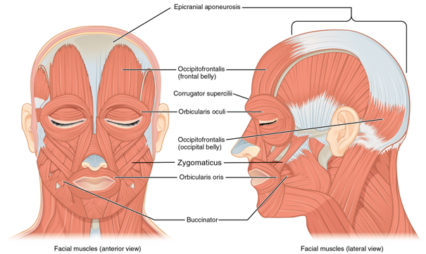
Facial Muscles
The orbicularis oris is a circular muscle that moves the lips and allows you to kiss. The orbicularis oculi is a circular muscle that closes the eye. The zygomaticus muscle pulls the lips up into a smile.
Chewing Muscles
In anatomical terminology, chewing is called mastication. Muscles involved in chewing must be able to exert enough pressure to bite through and then chew food before it is swallowed. The massetermuscle is the main muscle used for chewing because it elevates the mandible (lower jaw) to close the mouth, and it is assisted by the temporalismuscle, which retracts the mandible (Figure 20.6). You can feel the temporalis move by putting your fingers to your temple as you chew.
Neck Muscles
The head, attached to the top of the vertebral column, is balanced, moved, and rotated by the neck muscles. When these muscles act unilaterally, the head rotates. When they contract on both sides, the head flexes or extends. The major muscle that flexes (ex- the head flexes when you look down) and rotates the head is the sternocleidomastoid (Figure 20.7). Place your fingers on both sides of the front of your neck and turn your head to the left and to the right. You will feel the movement originate there.
The trapezius muscle is located on the back of the neck, and it extends the head (ex- the head extends when you look up).
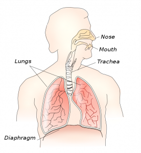
Thoracic Cavity Muscles
The muscles of the chest serve to facilitate breathing by changing the size of the thoracic cavity. When you inhale, your chest rises because the cavity expands. Alternately, when you exhale, your chest falls because the thoracic cavity decreases in size.
The change in volume of the thoracic cavity during breathing is due to the alternate contraction and relaxation of the diaphragm (Figures 20.8 and 20.9). It separates the thoracic and abdominal cavities, and is dome-shaped at rest. The superior surface of the diaphragm is convex, creating the elevated floor of the thoracic cavity. The inferior surface is concave, creating the curved roof of the abdominal cavity.
The external intercostal (inter- means between and -costal means rib) muscles are located in between the ribs (Figure 20.10). The principal role of the intercostal muscles is to assist in breathing by changing the dimensions of the rib cage. The external intercostal muscles aid in inspiration of air during breathing because when they contract, they raise the rib cage, which expands it.
Abdominal Wall Muscles
It is a complex job to balance the body on two feet and walk upright. The muscles of the vertebral column, thorax, and abdominal wall extend, flex, and stabilize different parts of the body’s trunk.
The rectus abdominis muscles (Figure 20.11) are a pair of long, linear muscles that extend the length of the abdomen. The rectus abdominis muscles flex the trunk (ex- when you do sit ups). Each muscle is segmented by three transverse bands of collagen fibers called the tendinous intersections. This results in the look of “six-pack abs,” as each segment hypertrophies on individuals at the gym who do many sit-ups.
On the lateral wall of the abdomen, the external oblique muscle is located closest to the surface, and the internal oblique muscle is underneath it (Figure 20.11). Both of these muscles flex the trunk laterally (bend to the side).
Shoulder Muscles
The pectoralis major is thick and fan-shaped, covering much of the top portion of the anterior (upper) thorax (Figure 20.12a). It flexes the shoulder (ex- throw a softball underhand or pick up a child). The deltoid is a thick muscle that creates the rounded lines of the shoulder (Figure 20.12b). The deltoid abducts the arm (the arm moves away from the body as when you reach out to the side).
Arm Muscles
The biceps brachii is located on the anterior (front) side of the arm and crosses the shoulder and elbow joints to flex the elbow and forearm (Figure 20.13). The triceps brachii is located on the posterior (back) side of the arm, and it extends the elbow and forearm (Figure 20.13).
Forearm Muscles
Their are several different forearm flexor and extensor muscles, but we will group them together because they have the same actions. The forearm flexors flex the wrist and fingers while the forearm extensors extend the wrist and fingers (Figure 20.13).
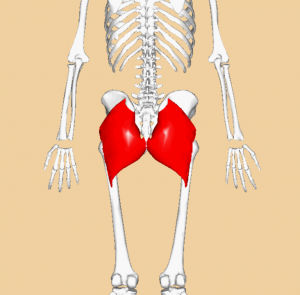
Hip Muscles
The psoas major and iliacus are located on the anterior (front) side of the hip and make up the iliopsoas muscle (Figure 20.14), which flexes the hip (ex- when you move your leg forward to kick a ball or when you lift your leg up to go up a step). The gluteus maximus is located on the posterior (back) side of the hip and makes up your butt (Figure 20.14 and 20.15). It extends the hip (ex- when you swing your leg back behind you).
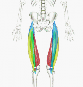
Thigh Muscles
Located on the anterior (front) side of the thigh, the quadriceps consists of four muscles (rectus femoris, vastus lateralis, vastus medialis, and vastus intermedius) (Figures 20.14 and 20.16). The tendon common to all four is the quadriceps tendon (patellar tendon), which inserts into the patella and continues below it as the patellar ligament. The patellar ligament attaches to the tibia. The quadriceps extends the knee (Figure 20.17).
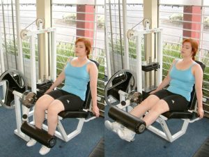
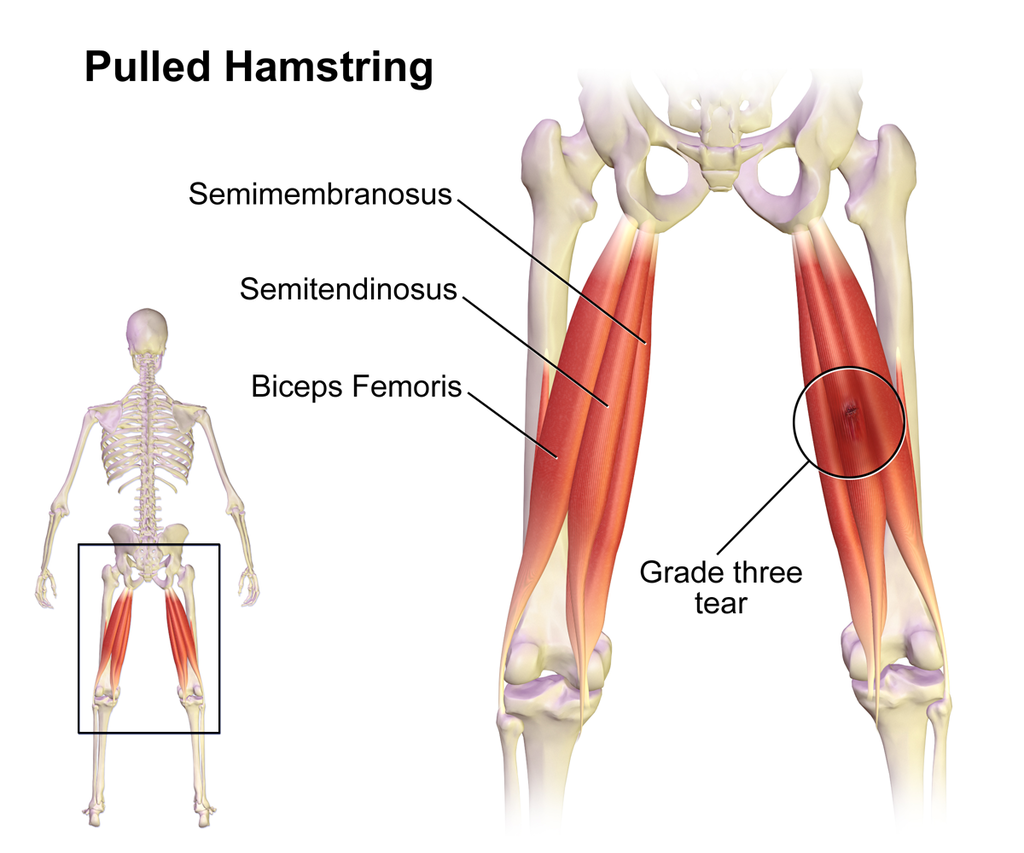
On the posterior (back) side of the thigh, the hamstring includes three long muscles (biceps femoris, semitendinosus, and semimembranosus) (Figures 20.14 and 20.18). The hamstrings flex the knee (Figure 20.17).
Lower Leg (Calf) Muscles
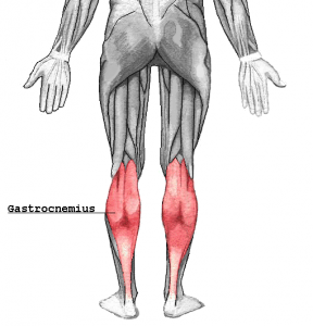
The tibialis anterior is a long and thick muscle on the lateral surface of the tibia (Figure 20.19) that flexes the ankle (ex- when you point your toes up towards your knee). On the posterior (back) side of the calf, the most superficial and visible muscle of the calf is the gastrocnemius (Figures 20.19 and 20.20). The gastrocnemius muscle extends the ankle (ex- when you point your toes down).
Adapted from Openstax Human Biology and Anatomy and Physiology
Media Attributions
- Facial muscles
- Diaphragm
- Gluteus maximus
- Quadriceps
- Quadriceps extends knee
- Hamstring
- Gastrocnemius
