
4 Chapter 4: Cell Structure and Transport
Chapter Outline
- 4.1 Studying Cells
- 4.2 Prokaryotic Cells
- 4.3 Eurkaryotic Cells
- 4.4 The Endomembrane System and Proteins
- 4.5 The Cytoskeleton
- 4.6 Connections between Cells and Cellular Activities
- 4.7 Plasma Membrane Components and Structure
- 4.8 Passive Transport
- 4.9 Active Transport
- 4.10 Bulk Transport
Close your eyes and picture a brick wall. What is the basic building block of that wall? A single brick, of course. Like a brick wall, your body is composed of basic building blocks, and the building blocks of your body are cells. Your body has many kinds of cells, each specialized for a specific purpose. Just as a home is made from a variety of building materials, the human body is constructed from many cell types. For example, epithelial cells protect the surface of the body and cover the organs and body cavities within. Bone cells help to support and protect the body. Cells of the immune system fight invading bacteria. Additionally, blood and blood cells carry nutrients and oxygen throughout the body while removing carbon dioxide. Each of these cell types plays a vital role during the growth, development, and day-to-day maintenance of the body. In spite of their enormous variety, however, cells from all organisms—even ones as diverse as bacteria, onion, and human—share certain fundamental characteristics.
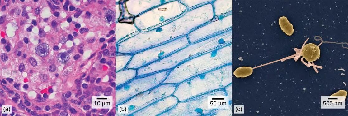
Figure 4.1 (a) Nasal sinus cells (viewed with a light microscope), (b) onion cells (viewed with a light microscope), and (c) Vibrio tasmaniensis bacterial cells (seen through a scanning electron microscope) are from very different organisms, yet all share certain characteristics of basic cell structure. (credit a: modification of work by Ed Uthman, MD; credit b: modification of work by Umberto Salvagnin; credit c: modification of work by Anthony D’Onofrio, William H. Fowle, Eric J. Stewart, and Kim Lewis of the Lewis Lab at Northeastern University; scale-bar data from Matt Russell)
Learning Objectives
You will be able to describe the basics of cells:
- Identify major cell components
- Differentiate the functions of cell part
- Be able to describe these characteristics of cellular transport:
- compare and contrast the different forms of membrane transport
- diffusion
- passive transport
- active transport
- vesicular transport
4.1 | Studying Cells
A cell is the smallest unit of a living thing. A living thing, whether made of one cell (like bacteria) or many cells (like a human), is called an organism. Thus, cells are the basic building blocks of all organisms. Several cells of one kind that interconnect with each other and perform a shared function form tissues, several tissues combine to form an organ (your stomach, heart, or brain), and several organs make up an organ system (such as the digestive system, circulatory system, or nervous system). Several systems that function together form an organism (like a human being). Here, we will examine the structure and function of cells. There are many types of cells, all grouped into one of two broad categories: prokaryotic and eukaryotic. For example, both animal and plant cells are classified as eukaryotic cells, whereas bacterial cells are classified as prokaryotic. Before discussing the criteria for determining whether a cell is prokaryotic or eukaryotic, let’s first examine how biologists study cells.
Microscopy
Cells vary in size. With few exceptions, individual cells cannot be seen with the naked eye, so scientists use microscopes (micro- = “small”; -scope = “to look at”) to study them. A microscope is an instrument that magnifies an object. Most photographs of cells are taken with a microscope, and these images can also be called micrographs.The optics of a microscope’s lenses change the orientation of the image that the user sees. A specimen that is right-side up and facing right on the microscope slide will appear upside-down and facing left when viewed through a microscope, and vice versa. Similarly, if the slide is moved left while looking through the microscope, it will appear to move right, and if moved down, it will seem to move up. This occurs because microscopes use two sets of lenses to magnify the image. Because of the manner by which light travels through the lenses, this system of two lenses produces an inverted image (binocular, or dissecting microscopes, work in a similar manner, but include an additional magnification system that makes the final image appear to be upright).
Light Microscopes
To give you a sense of cell size, a typical human red blood cell is about eight millionths of a meter or eight micrometers (abbreviated as eight μm) in diameter; the head of a pin of is about two thousandths of a meter (two mm) in diameter. That means about 250 red blood cells could fit on the head of a pin.Most student microscopes are classified as light microscopes (Figure 4.2a). Visible light passes and is bent through the lens system to enable the user to see the specimen. Light microscopes are advantageous for viewing living organisms, but since individual cells are generally transparent, their components are not distinguishable unless they are colored with special stains. Staining, however, usually kills the cells.Light microscopes commonly used in the undergraduate college laboratory magnify up to approximately 400 times. Two parameters that are important in microscopy are magnification and resolving power. Magnification is the process of enlarging an object in appearance. Resolving power is the ability of a microscope to distinguish two adjacent structures as separate: the higher the resolution, the better the clarity and detail of the image. When oil immersion lenses are used for the study of small objects, magnification is usually increased to 1,000 times. In order to gain a better understanding of cellular structure and function, scientists typically use electron microscopes.
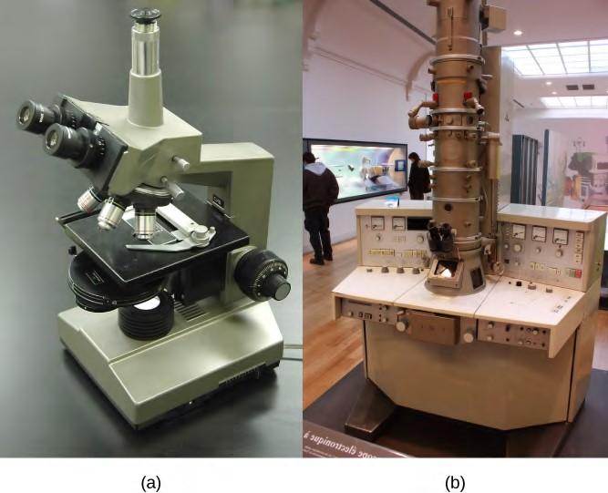
Figure 4.2 (a) Most light microscopes used in a college biology lab can magnify cells up to approximately 400 times and have a resolution of about 200 nanometers. (b) Electron microscopes provide a much higher magnification, 100,000x, and a have a resolution of 50 picometers. (credit a: modification of work by “GcG”/Wikimedia Commons; credit b: modification of work by Evan Bench)
Electron Microscopes
In contrast to light microscopes, electron microscopes (Figure 4.2b) use a beam of electrons instead of a beam of light. Not only does this allow for higher magnification and, thus, more detail (Figure 4.3), it also provides higher resolving power. The method used to prepare the specimen for viewing with an electron microscope kills the specimen. Electrons have short wavelengths (shorter than photons) that move best in a vacuum, so living cells cannot be viewed with an electron microscope. In a scanning electron microscope, a beam of electrons moves back and forth across a cell’s surface, creating details of cell surface characteristics. In a transmission electron microscope, the electron beam penetrates the cell and provides details of a cell’s internal structures. As you might imagine, electron microscopes are significantly more bulky and expensive than light microscopes.

(a)(b)
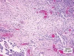
Figure 4.3 (a) These Salmonella bacteria appear as tiny purple dots when viewed with a light microscope. (b) This scanning electron microscope micrograph shows Salmonella bacteria (in red) invading human cells (yellow). Even though subfigure (b) shows a different Salmonella specimen than subfigure (a), you can still observe the comparative increase in magnification and detail. (credit a: modification of work by CDC/Armed Forces Institute of Pathology, Charles N. Farmer, Rocky Mountain Laboratories; credit b: modification of work by NIAID, NIH; scale-bar data from Matt Russell)
For another perspective on cell size, try the How Big interactive at this site(http://openstaxcollege.org/l/cell_sizes) .
Cell Theory
The microscopes we use today are far more complex than those used in the 1600s by Antony van Leeuwenhoek, a Dutch shopkeeper who had great skill in crafting lenses. Despite the limitations of his now-ancient lenses, van Leeuwenhoek observed the movements of protista (a type of single-celled organism) and sperm, which he collectively termed “animalcules.” In a 1665 publication called Micrographia, experimental scientist Robert Hooke coined the term “cell” for the box-like structures he observed when viewing cork tissue through a lens. In the 1670s, van Leeuwenhoek discovered bacteria and protozoa. Later advances in lenses, microscope construction, and staining techniques enabled other scientists to see some components inside cells. By the late 1830s, botanist Matthias Schleiden and zoologist Theodor Schwann were studying tissues and proposed the unified cell theory, which states that all living things are composed of one or more cells, the cell is the basic unit of life, and new cells arise from existing cells. Rudolf Virchow later made important contributions to this theory.
4.2 | Prokaryotic Cells
Cells fall into one of two broad categories: prokaryotic and eukaryotic. Only the predominantly single celled organisms of the domains Bacteria and Archaea are classified as prokaryotes (pro- = “before”; -kary- = “nucleus”). Cells of animals, plants, fungi, and protists are all eukaryotes (ceu- = “true”) and are made up of eukaryotic cells.
Components of Prokaryotic Cells
All cells share four common components: 1) a plasma membrane, an outer covering that separates the cell’s interior from its surrounding environment; 2) cytoplasm, consisting of a jelly-like cytosol within the cell in which other cellular components are found; 3) DNA, the genetic material of the cell; and 4) ribosomes, which synthesize proteins. However, prokaryotes differ from eukaryotic cells in several ways. A prokaryote is a simple, mostly single-celled (unicellular) organism that lacks a nucleus, or any other membrane-bound organelle. We will shortly come to see that this is significantly different in eukaryotes. Prokaryotic DNA is found in a central part of the cell: the nucleoid (Figure 4.5).
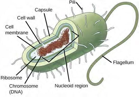
Figure 4.5 This figure shows the generalized structure of a prokaryotic cell. All prokaryotes have chromosomal DNA localized in a nucleoid, ribosomes, a cell membrane, and a cell wall. The other structures shown are present in some, but not all, bacteria.
Most prokaryotes have a peptidoglycan cell wall and many have a polysaccharide capsule (Figure 4.5). The cell wall acts as an extra layer of protection, helps the cell maintain its shape, and prevents dehydration. The capsule enables the cell to attach to surfaces in its environment. Some prokaryotes have flagella, pili, or fimbriae. Flagella are used for locomotion. Pili are used to exchange genetic material during a type of reproduction called conjugation. Fimbriae are used by bacteria to attach to a host cell.
Cell Size
At 0.1 to 5.0 μm in diameter, prokaryotic cells are significantly smaller than eukaryotic cells, which have diameters ranging from 10 to 100 μm (Figure 4.6). The small size of prokaryotes allows ions and organic molecules that enter them to quickly diffuse to other parts of the cell. Similarly, any wastes produced within a prokaryotic cell can quickly diffuse out. This is not the case in eukaryotic cells, which have developed different structural adaptations to enhance intracellular transport.
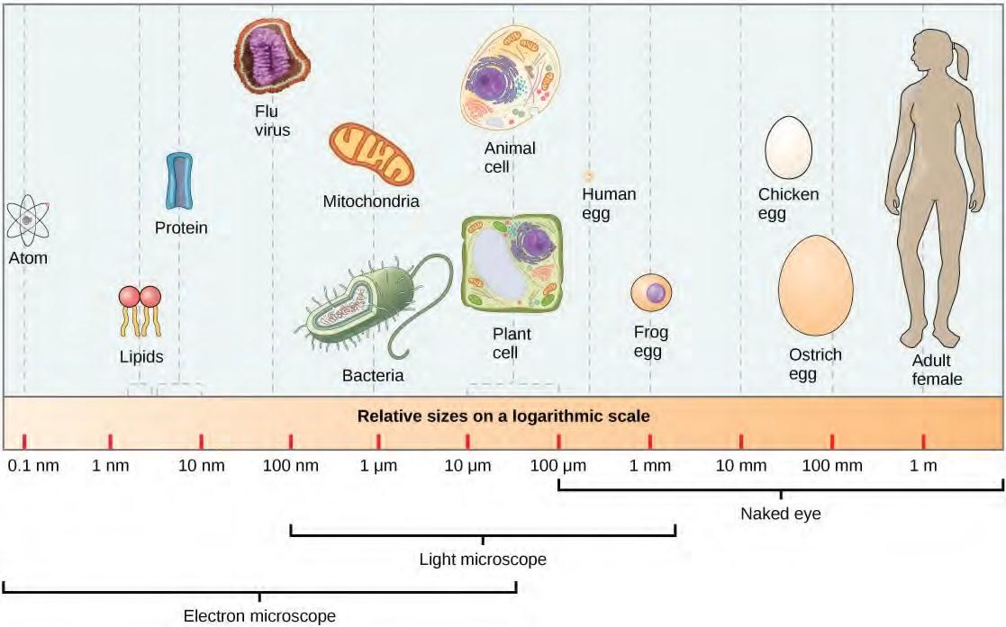
Figure 4.6 This figure shows relative sizes of microbes on a logarithmic scale (recall that each unit of increase in a logarithmic scale represents a 10-fold increase in the quantity being measured).
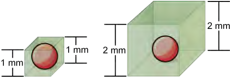
Small size, in general, is necessary for all cells, whether prokaryotic or eukaryotic. Let’s examine why that is so. First, we’ll consider the area and volume of a typical cell. Not all cells are spherical in shape, but most tend to approximate a sphere. You may remember from your high school geometry course that the formula for the surface area of a sphere is 4πr2, while the formula for its volume is 4πr3/3. Thus, as the radius of a cell increases, its surface area increases as the square of its radius, but its volume increases as the cube of its radius (much more rapidly). Therefore, as a cell increases in size, its surface area-to-volume ratio decreases. This same principle would apply if the cell had the shape of a cube (Figure 4.7). If the cell grows too large, the plasma membrane will not have sufficient surface area to support the rate of diffusion required for the increased volume. In other words, as a cell grows, it becomes less efficient. One way to become more efficient is to divide; another way is to develop organelles that perform specific tasks. These adaptations lead to the development of more sophisticated cells called eukaryotic cells.
4.3 | Eukaryotic Cells
Have you ever heard the phrase “form follows function?” It’s a philosophy practiced in many industries. In architecture, this means that buildings should be constructed to support the activities that will be carried out inside them. For example, a skyscraper should be built with several elevator banks; a hospital should be built so that its emergency room is easily accessible.
At this point, it should be clear to you that eukaryotic cells have a more complex structure than prokaryotic cells. Organelles allow different functions to be compartmentalized in different areas of the cell. Before turning to organelles, let’s first examine two important components of the cell: the plasma membrane and the cytoplasm.
The Plasma Membrane
Like prokaryotes, eukaryotic cells have a plasma membrane ( Figure 4.9), a phospholipid bilayer with embedded proteins that separates the internal contents of the cell from its surrounding environment. A phospholipid is a lipid molecule with two fatty acid chains and a phosphate-containing group. The plasma membrane controls the passage of organic molecules, ions, water, and oxygen into and out of the cell. Wastes (such as carbon dioxide and ammonia) also leave the cell by passing through the plasma membrane.
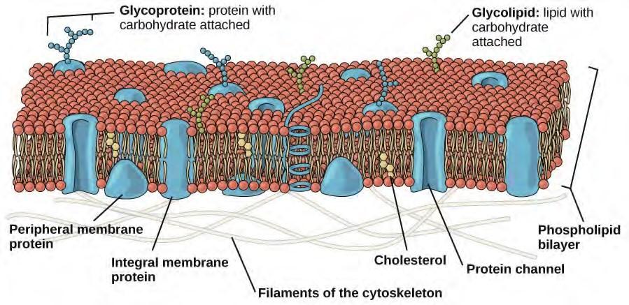
Figure 4.9 The eukaryotic plasma membrane is a phospholipid bilayer with proteins and cholesterol embedded in it.
The plasma membranes of cells that specialize in absorption are folded into fingerlike projections called microvilli (singular = microvillus); ( Figure 4.10). Such cells are typically found lining the small intestine, the organ that absorbs nutrients from digested food. This is an excellent example of form following function. People with celiac disease have an immune response to gluten, which is a protein found in wheat, barley, and rye. The immune response damages microvilli, and thus, afflicted individuals cannot absorb nutrients. This leads to malnutrition, cramping, and diarrhea. Patients suffering from celiac disease must follow a gluten-free diet.
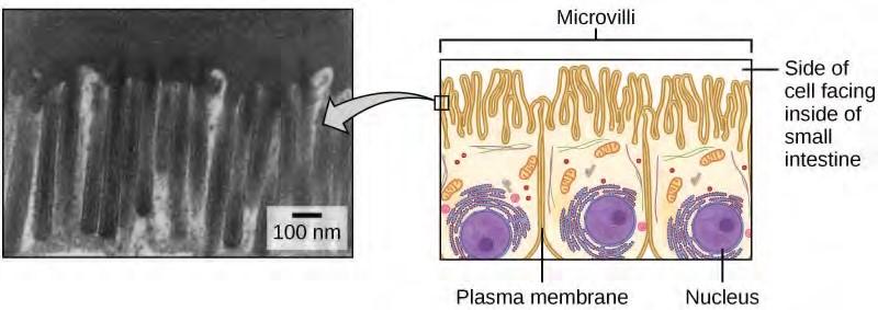
Figure 4.10 Microvilli, shown here as they appear on cells lining the small intestine, increase the surface area available for absorption. These microvilli are only found on the area of the plasma membrane that faces the cavity from which substances will be absorbed. (credit “micrograph”: modification of work by Louisa Howard)
The Cytoplasm
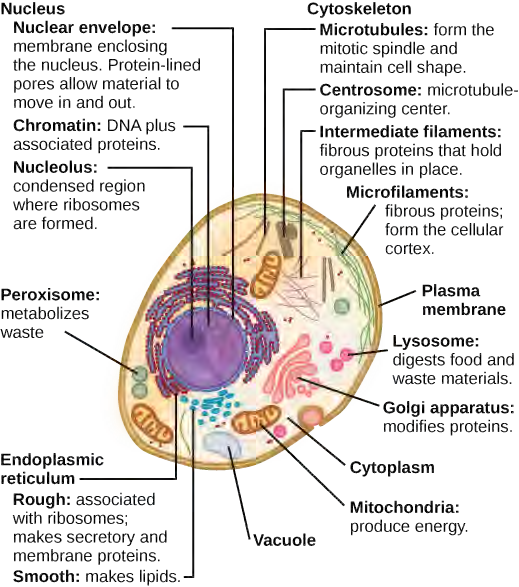
The cytoplasm is the entire region of a cell between the plasma membrane and the nuclear envelope (a structure to be discussed shortly). It is made up of organelles suspended in the gel-like cytosol, the cytoskeleton, and various chemicals ( Figure 4.8). Even though the cytoplasm consists of 70 to 80 percent water, it has a semi-solid consistency, which comes from the proteins within it. However, proteins are not the only organic molecules found in the cytoplasm. Glucose and other simple sugars, polysaccharides, amino acids, nucleic acids, fatty acids, and derivatives of glycerol are found there, too. Ions of sodium, potassium, calcium, and many other elements are also dissolved in the cytoplasm. Many metabolic reactions, including protein synthesis, take place in the cytoplasm.
The Nucleus
Typically, the nucleus is the most prominent organelle in a cell ( Figure 4.8). The nucleus (plural = nuclei) houses the cell’s DNA and directs the synthesis of ribosomes and proteins. Let’s look at it in more detail ( Figure 4.11).
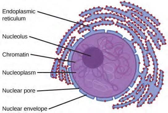
Figure 4.11 The nucleus stores chromatin (DNA plus proteins) in a gel-like substance called the nucleoplasm. The nucleolus is a condensed region of chromatin where ribosome synthesis occurs. The boundary of the nucleus is called the nuclear envelope. It consists of two phospholipid bilayers: an outer membrane and an inner membrane. The nuclear membrane is continuous with the endoplasmic reticulum. Nuclear pores allow substances to enter and exit the nucleus.
The Nuclear Envelope
The nuclear envelope is a double-membrane structure that constitutes the outermost portion of the nucleus ( Figure 4.11). Both the inner and outer membranes of the nuclear envelope are phospholipid bilayers.The nuclear envelope is punctuated with pores that control the passage of ions, molecules, and RNA between the nucleoplasm and cytoplasm. The nucleoplasm is the semi-solid fluid inside the nucleus, where we find the chromatin and the nucleolus.
Chromatin and Chromosomes
To understand chromatin, it is helpful to first consider chromosomes. Chromosomes are structures within the nucleus that are made up of DNA, the hereditary material. You may remember that in prokaryotes, DNA is organized into a single circular chromosome. In eukaryotes, chromosomes are linear structures. Every eukaryotic species has a specific number of chromosomes in the nuclei of its body’s cells. For example, in humans, the chromosome number is 46, while in fruit flies, it is eight. Chromosomes are only visible and distinguishable from one another when the cell is getting ready to divide. When the cell is in the growth and maintenance phases of its life cycle, proteins are attached to chromosomes, and they resemble an unwound, jumbled bunch of threads. These unwound protein chromosome complexes are called chromatin ( Figure 4.12); chromatin describes the material that makes up the chromosomes both when condensed and decondensed.
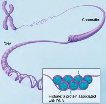

(a)(b)Figure 4.12 (a) This image shows various levels of the organization of chromatin (DNA and protein). (b) This image shows paired chromosomes. (credit b: modification of work by NIH; scale-bar data from Matt Russell)
The Nucleolus
We already know that the nucleus directs the synthesis of ribosomes, but how does it do this? Some chromosomes have sections of DNA that encode ribosomal RNA. A darkly staining area within the nucleus called the nucleolus (plural = nucleoli) aggregates the ribosomal RNA with associated proteins to assemble the ribosomal subunits that are then transported out through the pores in the nuclear envelope to the cytoplasm.
Ribosomes
Ribosomes are the cellular structures responsible for protein synthesis. When viewed through an electron microscope, ribosomes appear either as clusters (polyribosomes) or single, tiny dots that float freely in the cytoplasm. They may be attached to the cytoplasmic side of the plasma membrane or the cytoplasmic side of the endoplasmic reticulum and the outer membrane of the nuclear envelope ( Figure 4.8). Electron microscopy has shown us that ribosomes, which are large complexes of protein and RNA, consist of two subunits, aptly called large and small ( Figure 4.13). Ribosomes receive their “orders” for protein synthesis from the nucleus where the DNA is transcribed into messenger RNA (mRNA). The mRNA travels to the ribosomes, which translate the code provided by the sequence of the nitrogenous bases in the mRNA into a specific order of amino acids in a protein. Amino acids are the building blocks of proteins.
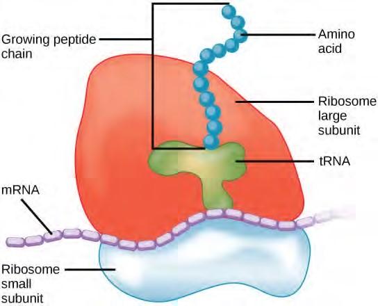
Figure 4.13 Ribosomes are made up of a large subunit (top) and a small subunit (bottom). During protein synthesis, ribosomes assemble amino acids into proteins.
Because protein synthesis is an essential function of all cells (including enzymes, hormones, antibodies, pigments, structural components, and surface receptors), ribosomes are found in practically every cell. Ribosomes are particularly abundant in cells that synthesize large amounts of protein. For example, the pancreas is responsible for creating several digestive enzymes and the cells that produce these enzymes contain many ribosomes. Thus, we see another example of form following function.
Mitochondria
Mitochondria (singular = mitochondrion) are often called the “powerhouses” or “energy factories” of a cell because they are responsible for making adenosine triphosphate (ATP), the cell’s main energy carrying molecule. ATP represents the short-term stored energy of the cell. Cellular respiration is the process of making ATP using the chemical energy found in glucose and other nutrients. In mitochondria, this process uses oxygen and produces carbon dioxide as a waste product. In fact, the carbon dioxide that you exhale with every breath comes from the cellular reactions that produce carbon dioxide as a byproduct.
Mitochondria are oval-shaped, double membrane organelles ( Figure 4.14) that have their own ribosomes and DNA. Each membrane is a phospholipid bilayer embedded with proteins. The inner layer has folds called cristae. The area surrounded by the folds is called the mitochondrial matrix. The cristae and the matrix have different roles in cellular respiration.
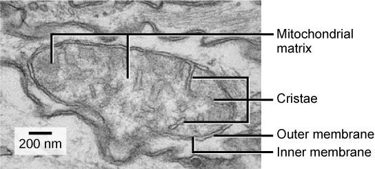
Figure 4.14 This electron micrograph shows a mitochondrion as viewed with a transmission electron microscope. This organelle has an outer membrane and an inner membrane. The inner membrane contains folds, called cristae, which increase its surface area. The space between the two membranes is called the intermembrane space, and the space inside the inner membrane is called the mitochondrial matrix. ATP synthesis takes place on the inner membrane. (credit: modification of work by Matthew Britton; scale-bar data from Matt Russell)
Peroxisomes
Peroxisomes are small, round organelles enclosed by single membranes. They carry out oxidation reactions that break down fatty acids and amino acids. They also detoxify many poisons that may enter the body. (Many of these oxidation reactions release hydrogen peroxide, H2O2, which would be damaging to cells; however, when these reactions are confined to peroxisomes, enzymes safely break down the H2O2 into oxygen and water.) For example, alcohol is detoxified by peroxisomes in liver cells. Glyoxysomes, which are specialized peroxisomes in plants, are responsible for converting stored fats into sugars.
Vesicles and Vacuoles
Vesicles and vacuoles are membrane-bound sacs that function in storage and transport. Other than the fact that vacuoles are somewhat larger than vesicles, there is a very subtle distinction between them: The membranes of vesicles can fuse with either the plasma membrane or other membrane systems within the cell. Additionally, some agents such as enzymes within plant vacuoles break down macromolecules. The membrane of a vacuole does not fuse with the membranes of other cellular components.
Animal Cells versus Plant Cells
At this point, you know that each eukaryotic cell has a plasma membrane, cytoplasm, a nucleus, ribosomes, mitochondria, peroxisomes, and in some, vacuoles, but there are some striking differences between animal and plant cells. While both animal and plant cells have microtubule organizing centers (MTOCs), animal cells also have centrioles associated with the MTOC: a complex called the centrosome. Animal cells each have a centrosome and lysosomes, whereas plant cells do not. Plant cells have a cell wall, chloroplasts and other specialized plastids, and a large central vacuole, whereas animal cells do not.
The Centrosome
The centrosome is a microtubule-organizing center found near the nuclei of animal cells. It contains a pair of centrioles, two structures that lie perpendicular to each other ( Figure 4.15). Each centriole is a cylinder of nine triplets of microtubules.
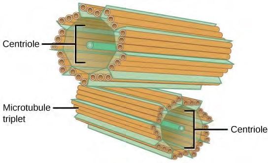
Figure 4.15 The centrosome consists of two centrioles that lie at right angles to each other. Each centriole is a cylinder made up of nine triplets of microtubules. Non-tubulin proteins (indicated by the green lines) hold the microtubule triplets together.
The centrosome (the organelle where all microtubules originate) replicates itself before a cell divides, and the centrioles appear to have some role in pulling the duplicated chromosomes to opposite ends of the dividing cell. However, the exact function of the centrioles in cell division isn’t clear, because cells that have had the centrosome removed can still divide, and plant cells, which lack centrosomes, are capable of cell division.
Lysosomes
Animal cells have another set of organelles not found in plant cells: lysosomes. The lysosomes are the cell’s “garbage disposal.” In plant cells, the digestive processes take place in vacuoles. Enzymes within the lysosomes aid the breakdown of proteins, polysaccharides, lipids, nucleic acids, and even worn-out organelles. These enzymes are active at a much lower pH than that of the cytoplasm. Therefore, the pH within lysosomes is more acidic than the pH of the cytoplasm. Many reactions that take place in the cytoplasm could not occur at a low pH, so again, the advantage of compartmentalizing the eukaryotic cell into organelles is apparent.
The Cell Wall
If you examine Figure 4.8b, the diagram of a plant cell, you will see a structure external to the plasma membrane called the cell wall. The cell wall is a rigid covering that protects the cell, provides structural support, and gives shape to the cell. Fungal and protistan cells also have cell walls. While the chief component of prokaryotic cell walls is peptidoglycan, the major organic molecule in the plant cell wall is cellulose ( Figure 4.16), a polysaccharide made up of glucose units. Have you ever noticed that when you bite into a raw vegetable, like celery, it crunches? That’s because you are tearing the rigid cell walls of the celery cells with your teeth.
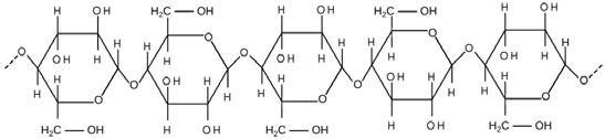
Figure 4.16 Cellulose is a long chain of β-glucose molecules connected by a 1-4 linkage. The dashed lines at each end of the figure indicate a series of many more glucose units. The size of the page makes it impossible to portray an entire cellulose molecule.
Chloroplasts
Like the mitochondria, chloroplasts have their own DNA and ribosomes, but chloroplasts have an entirely different function. Chloroplasts are plant cell organelles that carry out photosynthesis. Photosynthesis is the series of reactions that use carbon dioxide, water, and light energy to make glucose and oxygen. This is a major difference between plants and animals; plants (autotrophs) are able to make their own food, like sugars, while animals (heterotrophs) must ingest their food.
Like mitochondria, chloroplasts have outer and inner membranes, but within the space enclosed by a chloroplast’s inner membrane is a set of interconnected and stacked fluid-filled membrane sacs called thylakoids ( Figure 4.17). Each stack of thylakoids is called a granum (plural = grana). The fluid enclosed by the inner membrane that surrounds the grana is called the stroma.
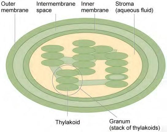
Figure 4.17 The chloroplast has an outer membrane, an inner membrane, and membrane structures called thylakoids that are stacked into grana. The space inside the thylakoid membranes is called the thylakoid space. The light harvesting reactions take place in the thylakoid membranes, and the synthesis of sugar takes place in the fluid inside the inner membrane, which is called the stroma. Chloroplasts also have their own genome, which is contained on a single circular chromosome.
The chloroplasts contain a green pigment called chlorophyll, which captures the light energy that drives the reactions of photosynthesis. Like plant cells, photosynthetic protists also have chloroplasts. Some bacteria perform photosynthesis, but their chlorophyll is not relegated to an organelle.
The Central Vacuole
Previously, we mentioned vacuoles as essential components of plant cells. If you look at Figure 4.8b, you will see that plant cells each have a large central vacuole that occupies most of the area of the cell. The central vacuole plays a key role in regulating the cell’s concentration of water in changing environmental conditions. Have you ever noticed that if you forget to water a plant for a few days, it wilts? That’s because as the water concentration in the soil becomes lower than the water concentration in the plant, water moves out of the central vacuoles and cytoplasm. As the central vacuole shrinks, it leaves the cell wall unsupported. This loss of support to the cell walls of plant cells results in the wilted appearance of the plant.
The central vacuole also supports the expansion of the cell. When the central vacuole holds more water, the cell gets larger without having to invest a lot of energy in synthesizing new cytoplasm.
4.4 | The Endomembrane System and Proteins
The endomembrane system (endo = “within”) is a group of membranes and organelles (Figure 4.18) in eukaryotic cells that works together to modify, package, and transport lipids and proteins. It includes the nuclear envelope, lysosomes, and vesicles, which we’ve already mentioned, and the endoplasmic reticulum and Golgi apparatus, which we will cover shortly. Although not technically within the cell, the plasma membrane is included in the endomembrane system because, as you will see, it interacts with the other endomembranous organelles. The endomembrane system does not include the membranes of either mitochondria or chloroplasts.
The Endoplasmic Reticulum
The endoplasmic reticulum (ER) (Figure 4.18) is a series of interconnected membranous sacs and tubules that collectively modifies proteins and synthesizes lipids. However, these two functions are performed in separate areas of the ER: the rough ER and the smooth ER, respectively.
The hollow portion of the ER tubules is called the lumen or cisternal space. The membrane of the ER, which is a phospholipid bilayer embedded with proteins, is continuous with the nuclear envelope.
Rough ER
The rough endoplasmic reticulum (RER) is so named because the ribosomes attached to its cytoplasmic surface give it a studded appearance when viewed through an electron microscope (Figure 4.19).
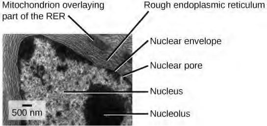
Figure 4.19 This transmission electron micrograph shows the rough endoplasmic reticulum and other organelles in a pancreatic cell. (credit: modification of work by Louisa Howard)
Ribosomes transfer their newly synthesized proteins into the lumen of the RER where they undergo structural modifications, such as folding or the acquisition of side chains. These modified proteins will be incorporated into cellular membranes—the membrane of the ER or those of other organelles—or secreted from the cell (such as protein hormones, enzymes). The RER also makes phospholipids for cellular membranes.
If the phospholipids or modified proteins are not destined to stay in the RER, they will reach their destinations via transport vesicles that bud from the RER’s membrane (Figure 4.18).
Since the RER is engaged in modifying proteins (such as enzymes, for example) that will be secreted from the cell, you would be correct in assuming that the RER is abundant in cells that secrete proteins.
This is the case with cells of the liver, for example.
Smooth ER
The smooth endoplasmic reticulum (SER) is continuous with the RER but has few or no ribosomes on its cytoplasmic surface (Figure 4.18). Functions of the SER include synthesis of carbohydrates, lipids, and steroid hormones; detoxification of medications and poisons; and storage of calcium ions.
In muscle cells, a specialized SER called the sarcoplasmic reticulum is responsible for storage of the calcium ions that are needed to trigger the coordinated contractions of the muscle cells.
You can watch an excellent animation of the endomembrane system here (http://openstaxcollege.org/ l/endomembrane) . At the end of the animation, there is a short self-assessment.
The Golgi Apparatus
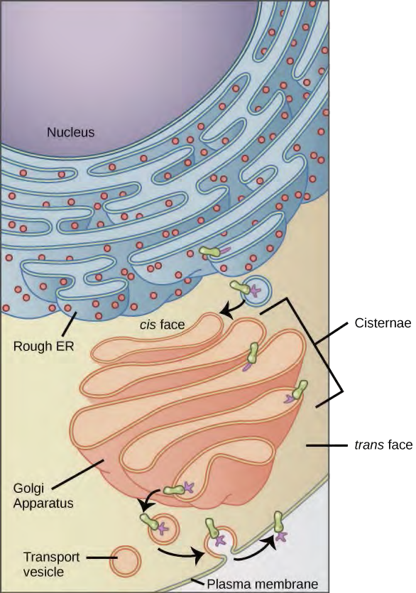
We have already mentioned that vesicles can bud from the ER and transport their contents elsewhere, but where do the vesicles go? Before reaching their final destination, the lipids or proteins within the transport vesicles still need to be sorted, packaged, and tagged so that they wind up in the right place. Sorting, tagging, packaging, and distribution of lipids and proteins takes place in the Golgi apparatus (also called the Golgi body), a series of flattened membranes (Figure 4.20).
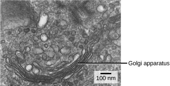
Figure 4.20 The Golgi apparatus in this white blood cell is visible as a stack of semicircular, flattened rings in the lower portion of the image. Several vesicles can be seen near the Golgi apparatus. (credit: modification of work by Louisa Howard)
The receiving side of the Golgi apparatus is called the cis face. The opposite side is called the trans face. The transport vesicles that formed from the ER travel to the cis face, fuse with it, and empty their contents into the lumen of the Golgi apparatus. As the proteins and lipids travel through the Golgi, they undergo further modifications that allow them to be sorted. The most frequent modification is the addition of short chains of sugar molecules. These newly modified proteins and lipids are then tagged with phosphate groups or other small molecules so that they can be routed to their proper destinations.
Finally, the modified and tagged proteins are packaged into secretory vesicles that bud from the trans face of the Golgi. While some of these vesicles deposit their contents into other parts of the cell where they will be used, other secretory vesicles fuse with the plasma membrane and release their contents outside the cell.
In another example of form following function, cells that engage in a great deal of secretory activity (such as cells of the salivary glands that secrete digestive enzymes or cells of the immune system that secrete antibodies) have an abundance of Golgi.
In plant cells, the Golgi apparatus has the additional role of synthesizing polysaccharides, some of which are incorporated into the cell wall and some of which are used in other parts of the cell.
Lysosomes
In addition to their role as the digestive component and organelle-recycling facility of animal cells, lysosomes are considered to be parts of the endomembrane system. Lysosomes also use their hydrolytic enzymes to destroy pathogens (disease-causing organisms) that might enter the cell. A good example of this occurs in a group of white blood cells called macrophages, which are part of your body’s immune system. In a process known as phagocytosis or endocytosis, a section of the plasma membrane of the macrophage invaginates (folds in) and engulfs a pathogen. The invaginated section, with the pathogen inside, then pinches itself off from the plasma membrane and becomes a vesicle. The vesicle fuses with a lysosome. The lysosome’s hydrolytic enzymes then destroy the pathogen (Figure 4.21).
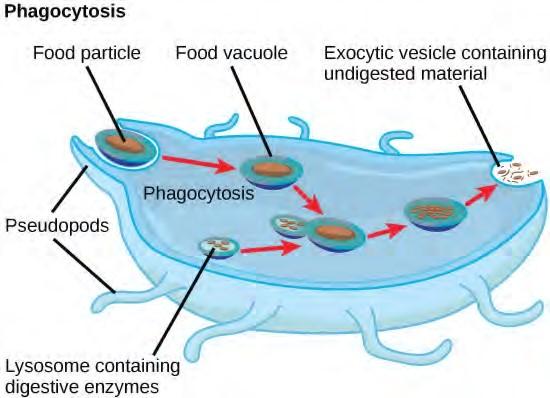
Figure 4.21 A macrophage has engulfed (phagocytized) a potentially pathogenic bacterium and then fuses with a lysosomes within the cell to destroy the pathogen. Other organelles are present in the cell but for simplicity are not shown.
4.5 | The Cytoskeleton
If you were to remove all the organelles from a cell, would the plasma membrane and the cytoplasm be the only components left? No. Within the cytoplasm, there would still be ions and organic molecules, plus a network of protein fibers that help maintain the shape of the cell, secure some organelles in specific positions, allow cytoplasm and vesicles to move within the cell, and enable cells within multicellular organisms to move. Collectively, this network of protein fibers is known as the cytoskeleton. There are three types of fibers within the cytoskeleton: microfilaments, intermediate filaments, and microtubules (Figure 4.22). Here, we will examine each.
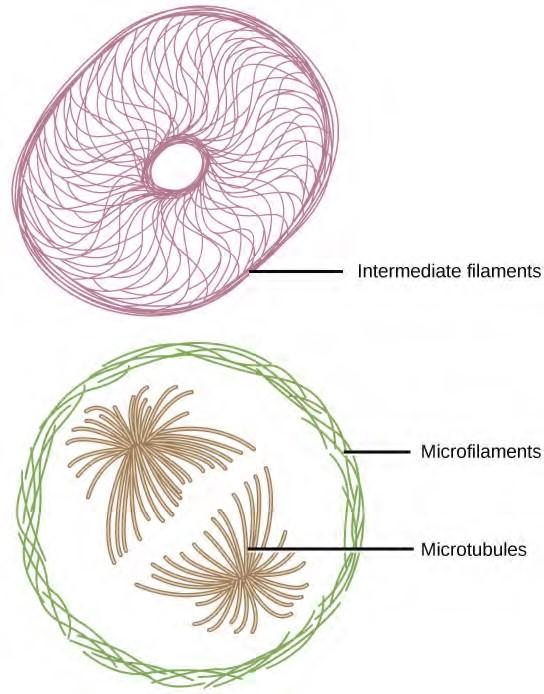
Figure 4.22 Microfilaments thicken the cortex around the inner edge of a cell; like rubber bands, they resist tension. Microtubules are found in the interior of the cell where they maintain cell shape by resisting compressive forces. Intermediate filaments are found throughout the cell and hold organelles in place.
Microfilaments
Of the three types of protein fibers in the cytoskeleton, microfilaments are the narrowest. They function in cellular movement, have a diameter of about 7 nm, and are made of two intertwined strands of a globular protein called actin (Figure 4.23). For this reason, microfilaments are also known as actin filaments.
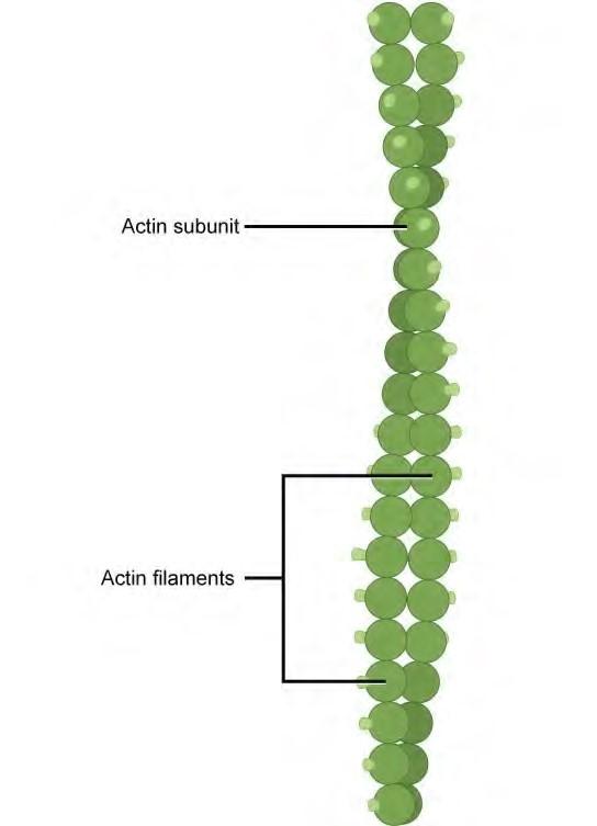
Figure 4.23 Microfilaments are made of two intertwined strands of actin.
Actin is powered by ATP to assemble its filamentous form, which serves as a track for the movement of a motor protein called myosin. This enables actin to engage in cellular events requiring motion, such as cell division in animal cells and cytoplasmic streaming, which is the circular movement of the cell cytoplasm in plant cells. Actin and myosin are plentiful in muscle cells. When your actin and myosin filaments slide past each other, your muscles contract.
Microfilaments also provide some rigidity and shape to the cell. They can depolymerize (disassemble) and reform quickly, thus enabling a cell to change its shape and move. White blood cells (your body’s infection-fighting cells) make good use of this ability. They can move to the site of an infection and phagocytize the pathogen.
To see an example of a white blood cell in action, click here (http://openstaxcollege.org/l/ chasing_bcteria) and watch a short time-lapse video of the cell capturing two bacteria. It engulfs one and then moves on to the other.
Intermediate Filaments
Intermediate filaments are made of several strands of fibrous proteins that are wound together (Figure 4.24). These elements of the cytoskeleton get their name from the fact that their diameter, 8 to 10 nm, is between those of microfilaments and microtubules.
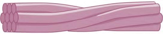
Figure 4.24 Intermediate filaments consist of several intertwined strands of fibrous proteins.
Intermediate filaments have no role in cell movement. Their function is purely structural. They bear tension, thus maintaining the shape of the cell, and anchor the nucleus and other organelles in place. Figure 4.22 shows how intermediate filaments create a supportive scaffolding inside the cell.
The intermediate filaments are the most diverse group of cytoskeletal elements. Several types of fibrous proteins are found in the intermediate filaments. You are probably most familiar with keratin, the fibrous protein that strengthens your hair, nails, and the epidermis of the skin.
Microtubules
As their name implies, microtubules are small hollow tubes. The walls of the microtubule are made of polymerized dimers of α-tubulin and β-tubulin, two globular proteins (Figure 4.25). With a diameter of about 25 nm, microtubules are the widest components of the cytoskeleton. They help the cell resist compression, provide a track along which vesicles move through the cell, and pull replicated chromosomes to opposite ends of a dividing cell. Like microfilaments, microtubules can dissolve and reform quickly.
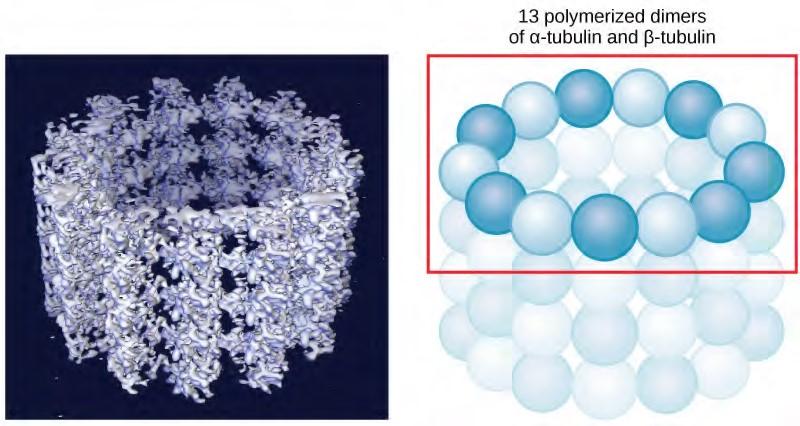
Figure 4.25 Microtubules are hollow. Their walls consist of 13 polymerized dimers of α-tubulin and β-tubulin (right image). The left image shows the molecular structure of the tube.
Microtubules are also the structural elements of flagella, cilia, and centrioles (the latter are the two perpendicular bodies of the centrosome). In fact, in animal cells, the centrosome is the microtubule organizing center. In eukaryotic cells, flagella and cilia are quite different structurally from their counterparts in prokaryotes, as discussed below.
Flagella and Cilia
To refresh your memory, flagella (singular = flagellum) are long, hair-like structures that extend from the plasma membrane and are used to move an entire cell (for example, sperm, Euglena). When present, the cell has just one flagellum or a few flagella. When cilia (singular = cilium) are present, however, many of them extend along the entire surface of the plasma membrane. They are short, hair-like structures that are used to move entire cells (such as paramecia) or substances along the outer surface of the cell (for example, the cilia of cells lining the Fallopian tubes that move the ovum toward the uterus, or cilia lining the cells of the respiratory tract that trap particulate matter and move it toward your nostrils.)
Despite their differences in length and number, flagella and cilia share a common structural arrangement of microtubules called a “9 + 2 array.” This is an appropriate name because a single flagellum or cilium is made of a ring of nine microtubule doublets, surrounding a single microtubule doublet in the center (Figure 4.26).
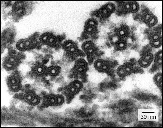
Figure 4.26 This transmission electron micrograph of two flagella shows the 9 + 2 array of microtubules: nine microtubule doublets surround a single microtubule doublet. (credit: modification of work by Dartmouth Electron Microscope Facility, Dartmouth College; scale-bar data from Matt Russell)
You have now completed a broad survey of the components of prokaryotic and eukaryotic cells. For a summary of cellular components in prokaryotic and eukaryotic cells, see Table 4.1.
Components of Prokaryotic and Eukaryotic Cells
|
Cell Component |
Present in Prokaryotes Function |
Present in Animal Cells? |
Present in Plant Cells? |
|
|
Plasma membrane |
Separates cell from external environment; controls passage of organic molecules, ions, water, oxygen, and wastes into and out of cell |
Yes |
Yes |
Yes |
|
Cytoplasm |
Provides turgor pressure to plant cells as fluid inside the central vacuole; site of many metabolic reactions; medium in which organelles are found |
Yes |
Yes |
Yes |
|
Nucleolus |
Darkened area within the nucleus where ribosomal subunits are synthesized. |
No |
Yes |
Yes |
|
Nucleus |
Cell organelle that houses DNA and directs synthesis of ribosomes and proteins |
No |
Yes |
Yes |
|
Ribosomes |
Protein synthesis |
Yes |
Yes |
Yes |
|
Mitochondria |
ATP production/cellular respiration |
No |
Yes |
Yes |
|
Peroxisomes |
Oxidizes and thus breaks down fatty acids and amino acids, and detoxifies poisons |
No |
Yes |
Yes |
|
Vesicles and vacuoles |
Storage and transport; digestive function in plant cells |
No |
Yes |
Yes |
|
Centrosome |
Unspecified role in cell division in animal cells; source of microtubules in animal cells |
No |
Yes |
No |
|
Lysosomes |
Digestion of macromolecules; recycling of worn-out organelles |
No |
Yes |
No |
|
Cell wall |
Protection, structural support and maintenance of cell shape |
Yes, primarily peptidoglycan |
No |
Yes, primarily cellulose |
|
Chloroplasts |
Photosynthesis |
No |
No |
Yes |
|
Endoplasmic reticulum |
Modifies proteins and synthesizes lipids |
No |
Yes |
Yes |
|
Golgi apparatus |
Modifies, sorts, tags, packages, and distributes lipids and proteins |
No |
Yes |
Yes |
Table 4.1
Components of Prokaryotic and Eukaryotic Cells
|
Cell Component |
Function |
Present in Prokaryotes? |
Present in Animal Cells? |
Present in Plant Cells? |
|
Cytoskeleton |
Maintains cell’s shape, secures organelles in specific positions, allows cytoplasm and vesicles to move within cell, and enables unicellular organisms to move independently |
Yes |
Yes |
Yes |
|
Flagella |
Cellular locomotion |
Some |
Some |
No, except for some plant sperm cells. |
|
Cilia |
Cellular locomotion, movement of particles along extracellular surface of plasma membrane, and filtration |
Some |
Some |
No |
Table 4.1
4.6 | Connections between Cells and Cellular Activities
You already know that a group of similar cells working together is called a tissue. As you might expect, if cells are to work together, they must communicate with each other, just as you need to communicate with others if you work on a group project. Let’s take a look at how cells communicate with each other.
Extracellular Matrix of Animal Cells
Most animal cells release materials into the extracellular space. The primary components of these materials are proteins, and the most abundant protein is collagen. Collagen fibers are interwoven with carbohydrate-containing protein molecules called proteoglycans. Collectively, these materials are called the extracellular matrix (Figure 4.27). Not only does the extracellular matrix hold the cells together to form a tissue, but it also allows the cells within the tissue to communicate with each other. How can this happen?
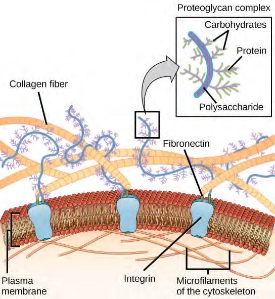
Figure 4.27 The extracellular matrix consists of a network of proteins and carbohydrates.
Cells have protein receptors on the extracellular surfaces of their plasma membranes. When a molecule within the matrix binds to the receptor, it changes the molecular structure of the receptor. The receptor, in turn, changes the conformation of the microfilaments positioned just inside the plasma membrane. These conformational changes induce chemical signals inside the cell that reach the nucleus and turn “on” or “off” the transcription of specific sections of DNA, which affects the production of associated proteins, thus changing the activities within the cell.
Blood clotting provides an example of the role of the extracellular matrix in cell communication. When the cells lining a blood vessel are damaged, they display a protein receptor called tissue factor. When tissue factor binds with another factor in the extracellular matrix, it causes platelets to adhere to the wall of the damaged blood vessel, stimulates the adjacent smooth muscle cells in the blood vessel to contract (thus constricting the blood vessel), and initiates a series of steps that stimulate the platelets to produce clotting factors.
Intercellular Junctions
Cells can also communicate with each other via direct contact, referred to as intercellular junctions. There are some differences in the ways that plant and animal cells do this. Plasmodesmata are junctions between plant cells, whereas animal cell contacts include tight junctions, gap junctions, and desmosomes.
Plasmodesmata
In general, long stretches of the plasma membranes of neighboring plant cells cannot touch one another because they are separated by the cell wall that surrounds each cell (Figure 4.8b). How then, can a plant transfer water and other soil nutrients from its roots, through its stems, and to its leaves? Such transport uses the vascular tissues (xylem and phloem) primarily. There also exist structural modifications called plasmodesmata (singular = plasmodesma), numerous channels that pass between cell walls of adjacent plant cells, connect their cytoplasm, and enable materials to be transported from cell to cell, and thus throughout the plant (Figure 4.28).
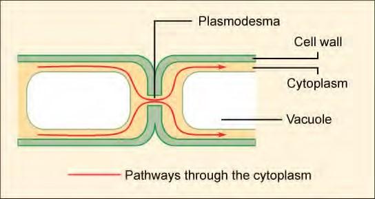
Figure 4.28 A plasmodesma is a channel between the cell walls of two adjacent plant cells. Plasmodesmata allow materials to pass from the cytoplasm of one plant cell to the cytoplasm of an adjacent cell.
Tight Junctions
A tight junction is a watertight seal between two adjacent animal cells (Figure 4.29). The cells are held tightly against each other by proteins (predominantly two proteins called claudins and occludins).
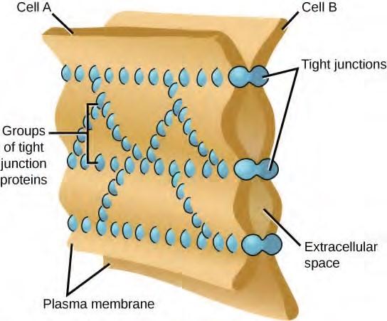
Figure 4.29 Tight junctions form watertight connections between adjacent animal cells. Proteins create tight junction adherence. (credit: modification of work by Mariana Ruiz Villareal)
This tight adherence prevents materials from leaking between the cells; tight junctions are typically found in epithelial tissues that line internal organs and cavities, and comprise most of the skin. For example, the tight junctions of the epithelial cells lining your urinary bladder prevent urine from leaking out into the extracellular space.
Desmosomes
Also found only in animal cells are desmosomes, which act like spot welds between adjacent epithelial cells (Figure 4.30). Short proteins called cadherins in the plasma membrane connect to intermediate filaments to create desmosomes. The cadherins join two adjacent cells together and maintain the cells in a sheet-like formation in organs and tissues that stretch, like the skin, heart, and muscles.
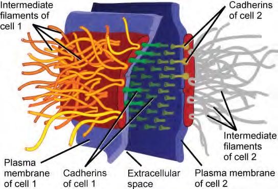
Figure 4.30 A desmosome forms a very strong spot weld between cells. It is created by the linkage
of cadherins and intermediate filaments. (credit: modification of work by Mariana Ruiz Villareal)
Gap Junctions
Gap junctions in animal cells are like plasmodesmata in plant cells in that they are channels between adjacent cells that allow for the transport of ions, nutrients, and other substances that enable cells to communicate (Figure 4.31). Structurally, however, gap junctions and plasmodesmata differ.
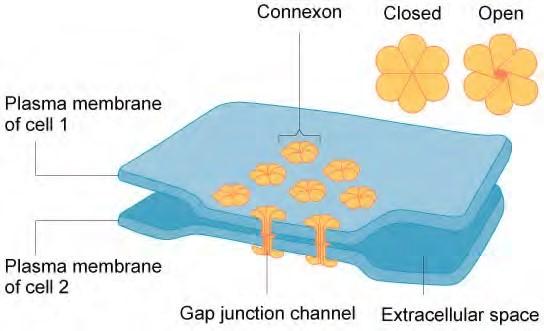
Figure 4.31 A gap junction is a protein-lined pore that allows water and small molecules to pass between adjacent animal cells. (credit: modification of work by Mariana Ruiz Villareal)
Gap junctions develop when a set of six proteins (called connexins) in the plasma membrane arrange themselves in an elongated donut-like configuration called a connexon. When the pores (“doughnut holes”) of connexons in adjacent animal cells align, a channel between the two cells forms. Gap junctions are particularly important in cardiac muscle: The electrical signal for the muscle to contract is passed efficiently through gap junctions, allowing the heart muscle cells to contract in tandem.
To conduct a virtual microscopy lab and review the parts of a cell, work through the steps of this interactive assignment (http://openstaxcollege.org/l/microscopy_lab) .

Figure 4.32 Despite its seeming hustle and bustle, Grand Central Station functions with a high level of organization: People and objects move from one location to another, they cross or are contained within certain boundaries, and they provide a constant flow as part of larger activity. Analogously, a plasma membrane’s functions involve movement within the cell and across boundaries in the process of intracellular and intercellular activities. (credit: modification of work by Randy Le’Moine)
4.7 | Plasma Membrane Components and Structure
A cell’s plasma membrane defines the cell, outlines its borders, and determines the nature of its interaction with its environment (see Table 4.2 for a summary). Cells exclude some substances, take in others, and excrete still others, all in controlled quantities. The plasma membrane must be very flexible to allow certain cells, such as red blood cells and white blood cells, to change shape as they pass through narrow capillaries. These are the more obvious functions of a plasma membrane. In addition, the surface of the plasma membrane carries markers that allow cells to recognize one another, which is vital for tissue and organ formation during early development, and which later plays a role in the “self” versus “non-self” distinction of the immune response.
Among the most sophisticated functions of the plasma membrane is the ability to transmit signals by means of complex, integral proteins known as receptors. These proteins act both as receivers of extracellular inputs and as activators of intracellular processes. These membrane receptors provide extracellular attachment sites for effectors like hormones and growth factors, and they activate intracellular response cascades when their effectors are bound. Occasionally, receptors are hijacked by viruses (HIV, human immunodeficiency virus, is one example) that use them to gain entry into cells, and at times, the genes encoding receptors become mutated, causing the process of signal transduction to malfunction with disastrous consequences.
Fluid Mosaic Model
The existence of the plasma membrane was identified in the 1890s, and its chemical components were identified in 1915. The principal components identified at that time were lipids and proteins. The first widely accepted model of the plasma membrane’s structure was proposed in 1935 by Hugh Davson and James Danielli; it was based on the “railroad track” appearance of the plasma membrane in early electron micrographs. They theorized that the structure of the plasma membrane resembles a sandwich, with protein being analogous to the bread, and lipids being analogous to the filling. In the 1950s, advances in microscopy, notably transmission electron microscopy (TEM), allowed researchers to see that the core of the plasma membrane consisted of a double, rather than a single, layer. A new model that better explains both the microscopic observations and the function of that plasma membrane was proposed by S.J. Singer and Garth L. Nicolson in 1972.
The explanation proposed by Singer and Nicolson is called the fluid mosaic model. The model has evolved somewhat over time, but it still best accounts for the structure and functions of the plasma membrane as we now understand them. The fluid mosaic model describes the structure of the plasma membrane as a mosaic of components—including phospholipids, cholesterol, proteins, and carbohydrates—that gives the membrane a fluid character. Plasma membranes range from 5 to 10 nm in thickness. For comparison, human red blood cells, visible via light microscopy, are approximately 8 µm wide, or approximately 1,000 times wider than a plasma membrane. The membrane does look a bit like a sandwich (Figure 4.33).
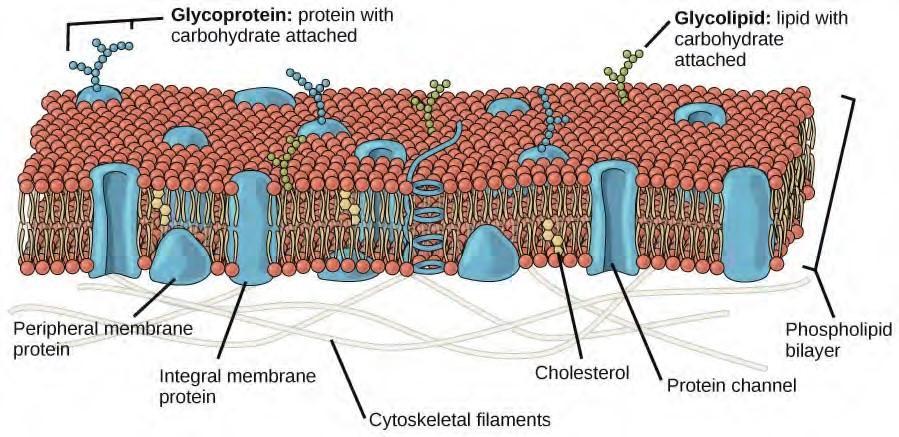
Figure 4.33 The fluid mosaic model of the plasma membrane describes the plasma membrane as a fluid combination of phospholipids, cholesterol, and proteins. Carbohydrates attached to lipids (glycolipids) and to proteins (glycoproteins) extend from the outward-facing surface of the membrane.
The principal components of a plasma membrane are lipids (phospholipids and cholesterol), proteins, and carbohydrates attached to some of the lipids and some of the proteins. A phospholipid is a molecule consisting of glycerol, two fatty acids, and a phosphate-linked head group. Cholesterol, another lipid composed of four fused carbon rings, is found alongside the phospholipids in the core of the membrane. The proportions of proteins, lipids, and carbohydrates in the plasma membrane vary with cell type, but for a typical human cell, protein accounts for about 50 percent of the composition by mass, lipids (of all types) account for about 40 percent of the composition by mass, with the remaining 10 percent of the composition by mass being carbohydrates. However, the concentration of proteins and lipids varies with different cell membranes. For example, myelin, an outgrowth of the membrane of specialized cells that insulates the axons of the peripheral nerves, contains only 18 percent protein and 76 percent lipid. The mitochondrial inner membrane contains 76 percent protein and only 24 percent lipid. The plasma membrane of human red blood cells is 30 percent lipid. Carbohydrates are present only on the exterior surface of the plasma membrane and are attached to proteins, forming glycoproteins, or attached to lipids, forming glycolipids.
Phospholipids
The main fabric of the membrane is composed of amphiphilic, phospholipid molecules. The hydrophilic or “water-loving” areas of these molecules (which look like a collection of balls in an artist’s rendition of the model) (Figure 4.33) are in contact with the aqueous fluid both inside and outside the cell. Hydrophobic, or water-hating molecules, tend to be non-polar. They interact with other non-polar molecules in chemical reactions, but generally do not interact with polar molecules. When placed in water, hydrophobic molecules tend to form a ball or cluster. The hydrophilic regions of the phospholipids tend to form hydrogen bonds with water and other polar molecules on both the exterior and interior of the cell. Thus, the membrane surfaces that face the interior and exterior of the cell are hydrophilic. In contrast, the interior of the cell membrane is hydrophobic and will not interact with water. Therefore, phospholipids form an excellent two-layer cell membrane that separates fluid within the cell from the fluid outside of the cell.
A phospholipid molecule (Figure 4.34) consists of a three-carbon glycerol backbone with two fatty acid molecules attached to carbons 1 and 2, and a phosphate-containing group attached to the third carbon. This arrangement gives the overall molecule an area described as its head (the phosphate-containing group), which has a polar character or negative charge, and an area called the tail (the fatty acids), which has no charge. The head can form hydrogen bonds, but the tail cannot. A molecule with this arrangement of a positively or negatively charged area and an uncharged, or non-polar, area is referred to as amphiphilic or “dual-loving.”
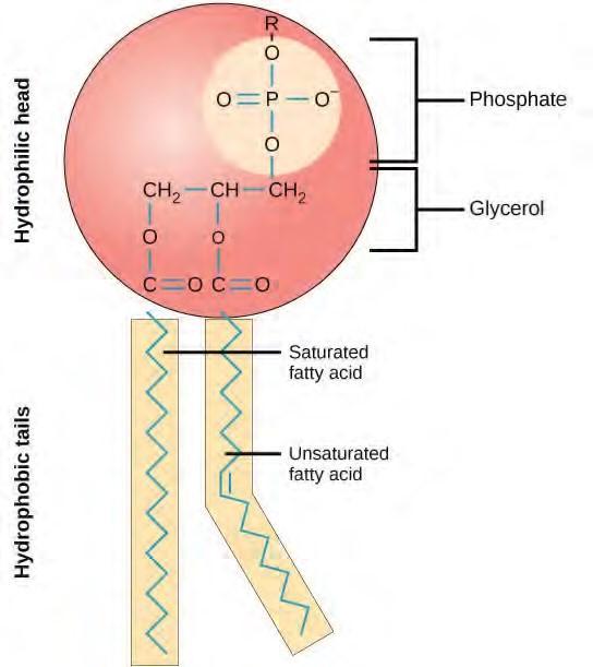
Figure 4.34 This phospholipid molecule is composed of a hydrophilic head and two hydrophobic tails. The hydrophilic head group consists of a phosphate-containing group attached to a glycerol molecule. The hydrophobic tails, each containing either a saturated or an unsaturated fatty acid, are long hydrocarbon chains.
This characteristic is vital to the structure of a plasma membrane because, in water, phospholipids tend to become arranged with their hydrophobic tails facing each other and their hydrophilic heads facing out. In this way, they form a lipid bilayer—a barrier composed of a double layer of phospholipids that separates the water and other materials on one side of the barrier from the water and other materials on the other side. In fact, phospholipids heated in an aqueous solution tend to spontaneously form small spheres or droplets (called micelles or liposomes), with their hydrophilic heads forming the exterior and their hydrophobic tails on the inside (Figure 4.35).
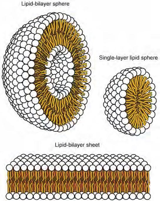
Figure 4.34 In an aqueous solution, phospholipids tend to arrange themselves with their polar heads facing outward and their hydrophobic tails facing inward. (credit: modification of work by MarianaRuiz Villareal)
Proteins
Proteins make up the second major component of plasma membranes. Integral proteins (some specialized types are called integrins) are, as their name suggests, integrated completely into the membrane structure, and their hydrophobic membrane-spanning regions interact with the hydrophobic region of the the phospholipid bilayer (Figure 4.33). Single-pass integral membrane proteins usually have a hydrophobic transmembrane segment that consists of 20–25 amino acids. Some span only part of the membrane—associating with a single layer—while others stretch from one side of the membrane to the other, and are exposed on either side. Some complex proteins are composed of up to 12 segments of a single protein, which are extensively folded and embedded in the membrane (Figure 4.36). This type of protein has a hydrophilic region or regions, and one or several mildly hydrophobic regions. This arrangement of regions of the protein tends to orient the protein alongside the phospholipids, with the hydrophobic region of the protein adjacent to the tails of the phospholipids and the hydrophilic region or regions of the protein protruding from the membrane and in contact with the cytosol or extracellular fluid.
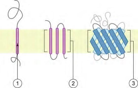
Figure 4.36 Integral membranes proteins may have one or more alpha-helices that span the membrane (examples 1 and 2), or they may have beta-sheets that span the membrane (example 3). (credit: “Foobar”/Wikimedia Commons)
Peripheral proteins are found on the exterior and interior surfaces of membranes, attached either to integral proteins or to phospholipids. Peripheral proteins, along with integral proteins, may serve as enzymes, as structural attachments for the fibers of the cytoskeleton, or as part of the cell’s recognition sites. These are sometimes referred to as “cell-specific” proteins. The body recognizes its own proteins and attacks foreign proteins associated with invasive pathogens.
Carbohydrates
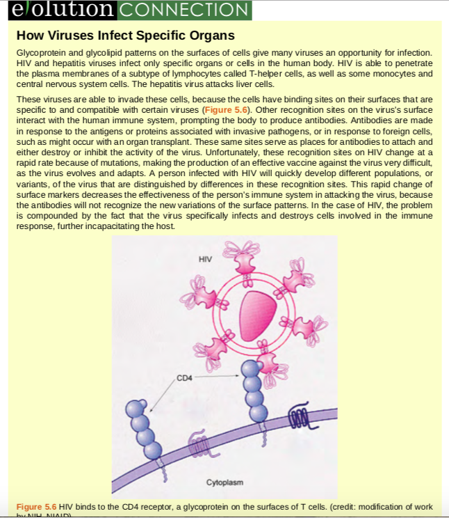
Carbohydrates are the third major component of plasma membranes. They are always found on the exterior surface of cells and are bound either to proteins (forming glycoproteins) or to lipids (forming glycolipids) (Figure 4.33). These carbohydrate chains may consist of 2–60 monosaccharide units and can be either straight or branched. Along with peripheral proteins, carbohydrates form specialized sites on the cell surface that allow cells to recognize each other. These sites have unique patterns that allow the cell to be recognized, much the way that the facial features unique to each person allow him or her to be recognized. This recognition function is very important to cells, as it allows the immune system to differentiate between body cells (called “self”) and foreign cells or tissues (called “non-self”). Similar types of glycoproteins and glycolipids are found on the surfaces of viruses and may change frequently, preventing immune cells from recognizing and attacking them.
These carbohydrates on the exterior surface of the cell—the carbohydrate components of both glycoproteins and glycolipids—are collectively referred to as the glycocalyx (meaning “sugar coating”). The glycocalyx is highly hydrophilic and attracts large amounts of water to the surface of the cell. This aids in the interaction of the cell with its watery environment and in the cell’s ability to obtain substances dissolved in the water. As discussed above, the glycocalyx is also important for cell identification, self/ non-self determination, and embryonic development, and is used in cell-cell attachments to form tissues
Membrane Fluidity
The mosaic characteristic of the membrane, described in the fluid mosaic model, helps to illustrate its nature. The integral proteins and lipids exist in the membrane as separate but loosely attached molecules. These resemble the separate, multicolored tiles of a mosaic picture, and they float, moving somewhat with respect to one another. The membrane is not like a balloon, however, that can expand and contract; rather, it is fairly rigid and can burst if penetrated or if a cell takes in too much water. However, because of its mosaic nature, a very fine needle can easily penetrate a plasma membrane without causing it to burst, and the membrane will flow and self-seal when the needle is extracted.
Thus, if saturated fatty acids, with their straight tails, are compressed by decreasing temperatures, they press in on each other, making a dense and fairly rigid membrane. If unsaturated fatty acids are compressed, the “kinks” in their tails elbow adjacent phospholipid molecules away, maintaining some space between the phospholipid molecules. This “elbow room” helps to maintain fluidity in the membrane at temperatures at which membranes with saturated fatty acid tails in their phospholipids would “freeze” or solidify. The relative fluidity of the membrane is particularly important in a cold environment. A cold environment tends to compress membranes composed largely of saturated fatty acids, making them less fluid and more susceptible to rupturing. Many organisms (fish are one example) are capable of adapting to cold environments by changing the proportion of unsaturated fatty acids in their membranes in response to the lowering of the temperature.
Visit this site (http://openstaxcollege.org/l/biological_memb) to see animations of the fluidity and mosaic quality of membranes.
Animals have an additional membrane constituent that assists in maintaining fluidity. Cholesterol, which lies alongside the phospholipids in the membrane, tends to dampen the effects of temperature on the membrane. Thus, this lipid functions as a buffer, preventing lower temperatures from inhibiting fluidity and preventing increased temperatures from increasing fluidity too much. Thus, cholesterol extends, in both directions, the range of temperature in which the membrane is appropriately fluid and consequently functional. Cholesterol also serves other functions, such as organizing clusters of transmembrane proteins into lipid rafts.
The Components and Functions of the Plasma Membrane
|
Component |
Location |
|
Phospholipid |
Main fabric of the membrane |
|
Cholesterol |
Attached between phospholipids and between the two phospholipid layers |
|
Integral proteins (for example, integrins) |
Embedded within the phospholipid layer(s). May or may not penetrate through both layers |
|
Peripheral proteins |
On the inner or outer surface of the phospholipid bilayer; not embedded within the phospholipids |
|
Carbohydrates (components of glycoproteins and glycolipids) |
Generally attached to proteins on the outside membrane layer |
Table 4.2
4.8 | Passive Transport
Plasma membranes must allow certain substances to enter and leave a cell, and prevent some harmful materials from entering and some essential materials from leaving. In other words, plasma membranes are selectively permeable—they allow some substances to pass through, but not others. If they were to lose this selectivity, the cell would no longer be able to sustain itself, and it would be destroyed. Some cells require larger amounts of specific substances than do other cells; they must have a way of obtaining these materials from extracellular fluids. This may happen passively, as certain materials move back and forth, or the cell may have special mechanisms that facilitate transport. Some materials are so important to a cell that it spends some of its energy, hydrolyzing adenosine triphosphate (ATP), to obtain these materials. Red blood cells use some of their energy doing just that. All cells spend the majority of their energy to maintain an imbalance of sodium and potassium ions between the interior and exterior of the cell.
The most direct forms of membrane transport are passive. Passive transport is a naturally occurring phenomenon and does not require the cell to exert any of its energy to accomplish the movement. In passive transport, substances move from an area of higher concentration to an area of lower concentration. A physical space in which there is a range of concentrations of a single substance is said to have a concentration gradient.
Selective Permeability
Plasma membranes are asymmetric: the interior of the membrane is not identical to the exterior of the membrane. In fact, there is a considerable difference between the array of phospholipids and proteins between the two leaflets that form a membrane. On the interior of the membrane, some proteins serve to anchor the membrane to fibers of the cytoskeleton. There are peripheral proteins on the exterior of the membrane that bind elements of the extracellular matrix. Carbohydrates, attached to lipids or proteins, are also found on the exterior surface of the plasma membrane. These carbohydrate complexes help the cell bind substances that the cell needs in the extracellular fluid. This adds considerably to the selective nature of plasma membranes (Figure 4.38).

Figure 4.38 The exterior surface of the plasma membrane is not identical to the interior surface of the same membrane.
Polar substances present problems for the membrane. While some polar molecules connect easily with the outside of a cell, they cannot readily pass through the lipid core of the plasma membrane. Additionally, while small ions could easily slip through the spaces in the mosaic of the membrane, their charge prevents them from doing so. Ions such as sodium, potassium, calcium, and chloride must have special means of penetrating plasma membranes. Simple sugars and amino acids also need help with transport across plasma membranes, achieved by various transmembrane proteins (channels).
Diffusion
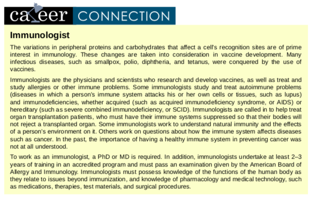
Diffusion is a passive process of transport. A single substance tends to move from an area of high concentration to an area of low concentration until the concentration is equal across a space. You are familiar with diffusion of substances through the air. For example, think about someone opening a bottle of ammonia in a room filled with people. The ammonia gas is at its highest concentration in the bottle; its lowest concentration is at the edges of the room. The ammonia vapor will diffuse, or spread away, from the bottle, and gradually, more and more people will smell the ammonia as it spreads. Materials move within the cell’s cytosol by diffusion, and certain materials move through the plasma membrane by diffusion (Figure 4.39). Diffusion expends no energy. On the contrary, concentration gradients are a form of potential energy, dissipated as the gradient is eliminated.
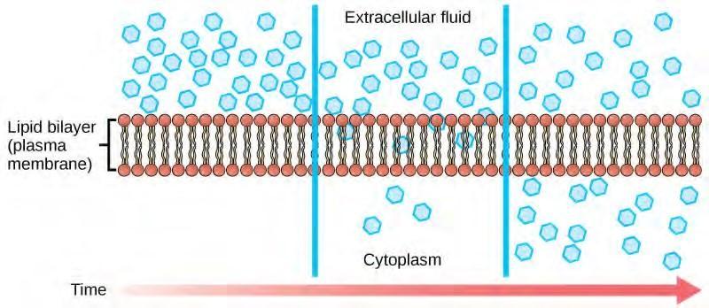
Figure 4.39 Diffusion through a permeable membrane moves a substance from an area of high concentration (extracellular fluid, in this case) down its concentration gradient (into the cytoplasm). (credit: modification of work by Mariana Ruiz Villareal)
Each separate substance in a medium, such as the extracellular fluid, has its own concentration gradient, independent of the concentration gradients of other materials. In addition, each substance will diffuse according to that gradient. Within a system, there will be different rates of diffusion of the different substances in the medium.
Factors That Affect Diffusion
Molecules move constantly in a random manner, at a rate that depends on their mass, their environment, and the amount of thermal energy they possess, which in turn is a function of temperature. This movement accounts for the diffusion of molecules through whatever medium in which they are localized. A substance will tend to move into any space available to it until it is evenly distributed throughout it. After a substance has diffused completely through a space, removing its concentration gradient, molecules will still move around in the space, but there will be no net movement of the number of molecules from one area to another. This lack of a concentration gradient in which there is no net movement of a substance is known as dynamic equilibrium. While diffusion will go forward in the presence of a concentration gradient of a substance, several factors affect the rate of diffusion.
- Extent of the concentration gradient: The greater the difference in concentration, the more rapid the diffusion. The closer the distribution of the material gets to equilibrium, the slower the rate of diffusion becomes.
- Mass of the molecules diffusing: Heavier molecules move more slowly; therefore, they diffuse more slowly. The reverse is true for lighter molecules.
- Temperature: Higher temperatures increase the energy and therefore the movement of the molecules, increasing the rate of diffusion. Lower temperatures decrease the energy of the molecules, thus decreasing the rate of diffusion.
- Solvent density: As the density of a solvent increases, the rate of diffusion decreases. The molecules slow down because they have a more difficult time getting through the denser medium. If the medium is less dense, diffusion increases. Because cells primarily use diffusion to move materials within the cytoplasm, any increase in the cytoplasm’s density will inhibit the movement of the materials. An example of this is a person experiencing dehydration. As the body’s cells lose water, the rate of diffusion decreases in the cytoplasm, and the cells’ functions deteriorate. Neurons tend to be very sensitive to this effect. Dehydration frequently leads to unconsciousness and possibly coma because of the decrease in diffusion rate within the cells.
- Solubility: As discussed earlier, nonpolar or lipid-soluble materials pass through plasma membranes more easily than polar materials, allowing a faster rate of diffusion.
- Surface area and thickness of the plasma membrane: Increased surface area increases the rate of diffusion, whereas a thicker membrane reduces it.
- Distance travelled: The greater the distance that a substance must travel, the slower the rate of diffusion. This places an upper limitation on cell size. A large, spherical cell will die because nutrients or waste cannot reach or leave the center of the cell, respectively. Therefore, cells must either be small in size, as in the case of many prokaryotes, or be flattened, as with many single celled eukaryotes.
A variation of diffusion is the process of filtration. In filtration, material moves according to its concentration gradient through a membrane; sometimes the rate of diffusion is enhanced by pressure, causing the substances to filter more rapidly. This occurs in the kidney, where blood pressure forces large amounts of water and accompanying dissolved substances, or solutes, out of the blood and into the renal tubules. The rate of diffusion in this instance is almost totally dependent on pressure. One of the effects of high blood pressure is the appearance of protein in the urine, which is “squeezed through” by the abnormally high pressure.
Facilitated transport
In facilitated transport, also called facilitated diffusion, materials diffuse across the plasma membrane with the help of membrane proteins. A concentration gradient exists that would allow these materials to diffuse into the cell without expending cellular energy. However, these materials are ions are polar molecules that are repelled by the hydrophobic parts of the cell membrane. Facilitated transport proteins shield these materials from the repulsive force of the membrane, allowing them to diffuse into the cell.
The material being transported is first attached to protein or glycoprotein receptors on the exterior surface of the plasma membrane. This allows the material that is needed by the cell to be removed from the extracellular fluid. The substances are then passed to specific integral proteins that facilitate their passage. Some of these integral proteins are collections of beta pleated sheets that form a pore or channel through the phospholipid bilayer. Others are carrier proteins which bind with the substance and aid its diffusion through the membrane.
Channels
The integral proteins involved in facilitated transport are collectively referred to as transport proteins, and they function as either channels for the material or carriers. In both cases, they are transmembrane proteins. Channels are specific for the substance that is being transported. Channel proteins have hydrophilic domains exposed to the intracellular and extracellular fluids; they additionally have a hydrophilic channel through their core that provides a hydrated opening through the membrane layers (Figure 4.40). Passage through the channel allows polar compounds to avoid the nonpolar central layer of the plasma membrane that would otherwise slow or prevent their entry into the cell. Aquaporins are channel proteins that allow water to pass through the membrane at a very high rate.
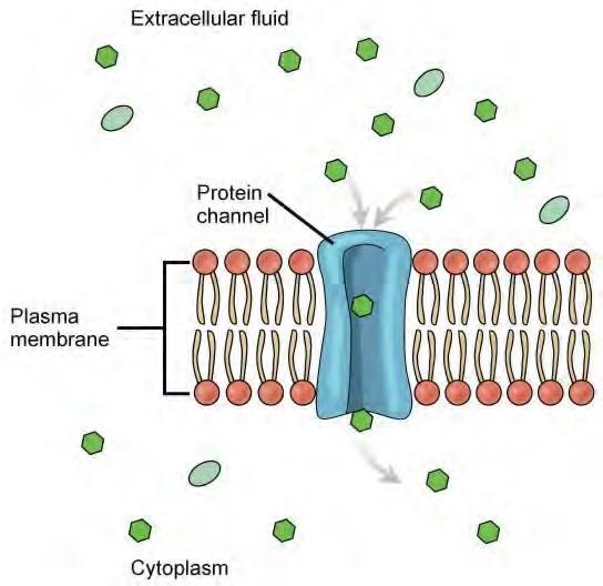
Figure 4.40 Facilitated transport moves substances down their concentration gradients. They may cross the plasma membrane with the aid of channel proteins. (credit: modification of work by MarianaRuiz Villareal)
Channel proteins are either open at all times or they are “gated,” which controls the opening of the channel. The attachment of a particular ion to the channel protein may control the opening, or other mechanisms or substances may be involved. In some tissues, sodium and chloride ions pass freely through open channels, whereas in other tissues a gate must be opened to allow passage. An example of this occurs in the kidney, where both forms of channels are found in different parts of the renal tubules. Cells involved in the transmission of electrical impulses, such as nerve and muscle cells, have gated channels for sodium, potassium, and calcium in their membranes. Opening and closing of these channels changes the relative concentrations on opposing sides of the membrane of these ions, resulting in the facilitation of electrical transmission along membranes (in the case of nerve cells) or in muscle contraction (in the case of muscle cells).
Carrier Proteins
Another type of protein embedded in the plasma membrane is a carrier protein. This aptly named protein binds a substance and, in doing so, triggers a change of its own shape, moving the bound molecule from the outside of the cell to its interior (Figure 4.41); depending on the gradient, the material may move in the opposite direction. Carrier proteins are typically specific for a single substance. This selectivity adds to the overall selectivity of the plasma membrane. The exact mechanism for the change of shape is poorly understood. Proteins can change shape when their hydrogen bonds are affected, but this may not fully explain this mechanism. Each carrier protein is specific to one substance, and there are a finite number of these proteins in any membrane. This can cause problems in transporting enough of the material for the cell to function properly. When all of the proteins are bound to their ligands, they are saturated and the rate of transport is at its maximum. Increasing the concentration gradient at this point will not result in an increased rate of transport.
Figure 4.41 Some substances are able to move down their concentration gradient across the plasma membrane with the aid of carrier proteins. Carrier proteins change shape as they move molecules across the membrane. (credit: modification of work by Mariana Ruiz Villareal)
An example of this process occurs in the kidney. Glucose, water, salts, ions, and amino acids needed by the body are filtered in one part of the kidney. This filtrate, which includes glucose, is then reabsorbed in another part of the kidney. Because there are only a finite number of carrier proteins for glucose, if more glucose is present than the proteins can handle, the excess is not transported and it is excreted from the body in the urine. In a diabetic individual, this is described as “spilling glucose into the urine.” A different group of carrier proteins called glucose transport proteins, or GLUTs, are involved in transporting glucose and other hexose sugars through plasma membranes within the body.
Channel and carrier proteins transport material at different rates. Channel proteins transport much more quickly than do carrier proteins. Channel proteins facilitate diffusion at a rate of tens of millions of molecules per second, whereas carrier proteins work at a rate of a thousand to a million molecules per second.
Osmosis
Osmosis is the movement of water through a semipermeable membrane according to the concentration gradient of water across the membrane, which is inversely proportional to the concentration of solutes. While diffusion transports material across membranes and within cells, osmosis transports only water across a membrane and the membrane limits the diffusion of solutes in the water. Not surprisingly, the aquaporins that facilitate water movement play a large role in osmosis, most prominently in red blood cells and the membranes of kidney tubules.
Mechanism
Osmosis is a special case of diffusion. Water, like other substances, moves from an area of high concentration to one of low concentration. An obvious question is what makes water move at all? Imagine a beaker with a semipermeable membrane separating the two sides or halves (Figure 4.42). On both sides of the membrane the water level is the same, but there are different concentrations of a dissolved substance, or solute, that cannot cross the membrane (otherwise the concentrations on each side would be balanced by the solute crossing the membrane). If the volume of the solution on both sides of the membrane is the same, but the concentrations of solute are different, then there are different amounts of water, the solvent, on either side of the membrane.
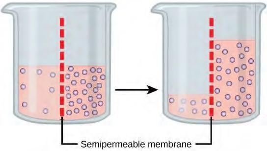
Figure 4.42 In osmosis, water always moves from an area of higher water concentration to one of lower concentration. In the diagram shown, the solute cannot pass through the selectively permeable membrane, but the water can.
Returning to the beaker example, recall that it has a mixture of solutes on either side of the membrane. A principle of diffusion is that the molecules move around and will spread evenly throughout the medium if they can. However, only the material capable of getting through the membrane will diffuse through it. In this example, the solute cannot diffuse through the membrane, but the water can. Water has a concentration gradient in this system. Thus, water will diffuse down its concentration gradient, crossing the membrane to the side where it is less concentrated. This diffusion of water through the membrane—osmosis—will continue until the concentration gradient of water goes to zero or until the hydrostatic pressure of the water balances the osmotic pressure. Osmosis proceeds constantly in living systems.
Tonicity
Tonicity describes how an extracellular solution can change the volume of a cell by affecting osmosis. A solution’s tonicity often directly correlates with the osmolarity of the solution. Osmolarity describes the total solute concentration of the solution. A solution with low osmolarity has a greater number of water molecules relative to the number of solute particles; a solution with high osmolarity has fewer water molecules with respect to solute particles. In a situation in which solutions of two different osmolarities are separated by a membrane permeable to water, though not to the solute, water will move from the side of the membrane with lower osmolarity (and more water) to the side with higher osmolarity (and less water). This effect makes sense if you remember that the solute cannot move across the membrane, and thus the only component in the system that can move—the water—moves along its own concentration gradient. An important distinction that concerns living systems is that osmolarity measures the number of particles (which may be molecules) in a solution. Therefore, a solution that is cloudy with cells may have a lower osmolarity than a solution that is clear, if the second solution contains more dissolved molecules than there are cells.
Hypotonic Solutions
Three terms—hypotonic, isotonic, and hypertonic—are used to relate the osmolarity of a cell to the osmolarity of the extracellular fluid that contains the cells. In a hypotonic situation, the extracellular fluid has lower osmolarity than the fluid inside the cell, and water enters the cell. (In living systems, the point of reference is always the cytoplasm, so the prefix hypo– means that the extracellular fluid has a lower concentration of solutes, or a lower osmolarity, than the cell cytoplasm.) It also means that the extracellular fluid has a higher concentration of water in the solution than does the cell. In this situation, water will follow its concentration gradient and enter the cell.
Hypertonic Solutions
As for a hypertonic solution, the prefix hyper– refers to the extracellular fluid having a higher osmolarity than the cell’s cytoplasm; therefore, the fluid contains less water than the cell does. Because the cell has a relatively higher concentration of water, water will leave the cell.
Isotonic Solutions
In an isotonic solution, the extracellular fluid has the same osmolarity as the cell. If the osmolarity of the cell matches that of the extracellular fluid, there will be no net movement of water into or out of the cell, although water will still move in and out. Blood cells and plant cells in hypertonic, isotonic, and hypotonic solutions take on characteristic appearances (Figure 4.43).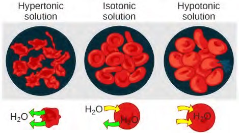
For a video illustrating the process of diffusion in solutions, visit this site (http://openstaxcollege.org/ l/dispersion) .
Tonicity in Living Systems
In a hypotonic environment, water enters a cell, and the cell swells. In an isotonic condition, the relative concentrations of solute and solvent are equal on both sides of the membrane. There is no net water movement; therefore, there is no change in the size of the cell. In a hypertonic solution, water leaves a cell and the cell shrinks. If either the hypo- or hyper- condition goes to excess, the cell’s functions become compromised, and the cell may be destroyed.
Various living things have ways of controlling the effects of osmosis—a mechanism called osmoregulation. Some organisms, such as plants, fungi, bacteria, and some protists, have cell walls that surround the plasma membrane and prevent cell lysis in a hypotonic solution. The plasma membrane can only expand to the limit of the cell wall, so the cell will not lyse. In fact, the cytoplasm in plants is always slightly hypertonic to the cellular environment, and water will always enter a cell if water is available. This inflow of water produces turgor pressure, which stiffens the cell walls of the plant (Figure 4.44). In nonwoody plants, turgor pressure supports the plant. Conversely, if the plant is not watered, the extracellular fluid will become hypertonic, causing water to leave the cell. In this condition, the cell does not shrink because the cell wall is not flexible. However, the cell membrane detaches from the wall and constricts the cytoplasm. This is called plasmolysis. Plants lose turgor pressure in this condition and wilt (Figure 4.46).
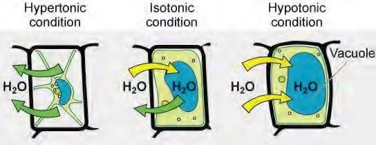
Figure 4.44 The turgor pressure within a plant cell depends on the tonicity of the solution that it is bathed in. (credit: modification of work by Mariana Ruiz Villareal)
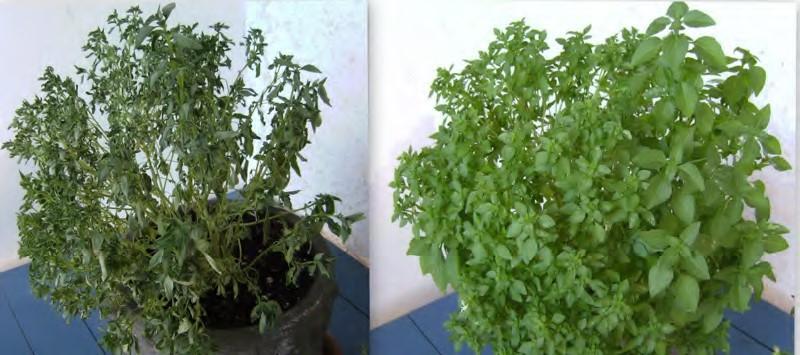
Figure 4.45 Without adequate water, the plant on the left has lost turgor pressure, visible in its wilting; the turgor pressure is restored by watering it (right). (credit: Victor M. Vicente Selvas)
Tonicity is a concern for all living things. For example, paramecia and amoebas, which are protists that lack cell walls, have contractile vacuoles. This vesicle collects excess water from the cell and pumps it out, keeping the cell from lysing as it takes on water from its environment (Figure 4.46).
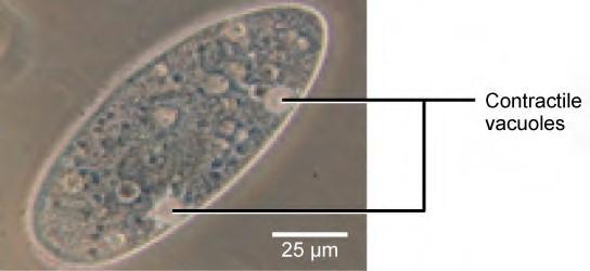
Figure 4.46 A paramecium’s contractile vacuole, here visualized using bright field light microscopy at 480x magnification, continuously pumps water out of the organism’s body to keep it from bursting in a hypotonic medium. (credit: modification of work by NIH; scale-bar data from Matt Russell)
Many marine invertebrates have internal salt levels matched to their environments, making them isotonic with the water in which they live. Fish, however, must spend approximately five percent of their metabolic energy maintaining osmotic homeostasis. Freshwater fish live in an environment that is hypotonic to their cells. These fish actively take in salt through their gills and excrete diluted urine to rid themselves of excess water. Saltwater fish live in the reverse environment, which is hypertonic to their cells, and they secrete salt through their gills and excrete highly concentrated urine.In vertebrates, the kidneys regulate the amount of water in the body. Osmoreceptors are specialized cells in the brain that monitor the concentration of solutes in the blood. If the levels of solutes increase beyond a certain range, a hormone is released that retards water loss through the kidney and dilutes the blood to safer levels. Animals also have high concentrations of albumin, which is produced by the liver, in their blood. This protein is too large to pass easily through plasma membranes and is a major factor in controlling the osmotic pressures applied to tissues.
4.9 | Active Transport
Active transport mechanisms require the use of the cell’s energy, usually in the form of adenosine triphosphate (ATP). If a substance must move into the cell against its concentration gradient—that is, if the concentration of the substance inside the cell is greater than its concentration in the extracellular fluid (and vice versa)—the cell must use energy to move the substance. Some active transport mechanisms move small-molecular weight materials, such as ions, through the membrane. Other mechanisms transport much larger molecules.
Electrochemical Gradient
We have discussed simple concentration gradients—differential concentrations of a substance across a space or a membrane—but in living systems, gradients are more complex. Because ions move into and out of cells and because cells contain proteins that do not move across the membrane and are mostly negatively charged, there is also an electrical gradient, a difference of charge, across the plasma membrane. The interior of living cells is electrically negative with respect to the extracellular fluid in which they are bathed, and at the same time, cells have higher concentrations of potassium (K+) and lower concentrations of sodium (Na+) than does the extracellular fluid. So in a living cell, the concentration gradient of Na+ tends to drive it into the cell, and the electrical gradient of Na+ (a positive ion) also tends to drive it inward to the negatively charged interior. The situation is more complex, however, for other elements such as potassium. The electrical gradient of K+, a positive ion, also tends to drive it into the cell, but the concentration gradient of K+ tends to drive K+ out of the cell (Figure 4.47). The combined gradient of concentration and electrical charge that affects an ion is called its electrochemical gradient.
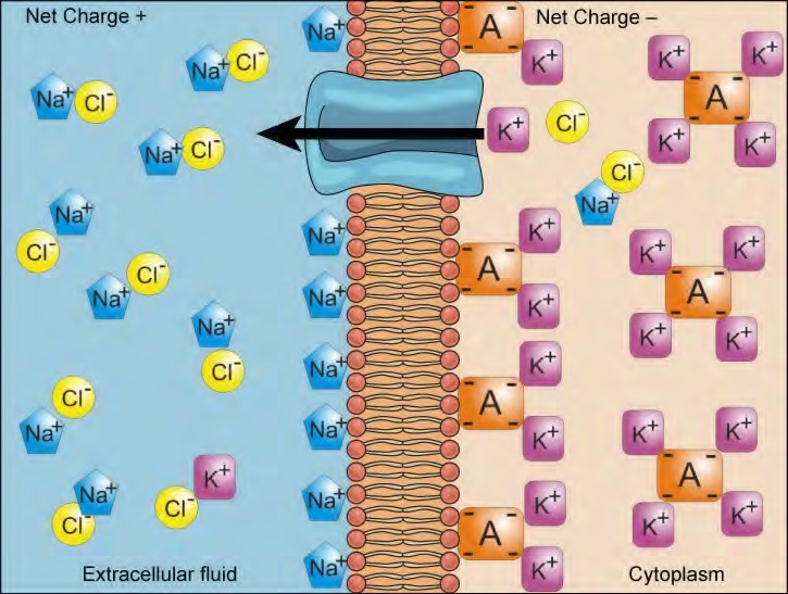
Moving Against a Gradient
To move substances against a concentration or electrochemical gradient, the cell must use energy. This energy is harvested from ATP generated through the cell’s metabolism. Active transport mechanisms, collectively called pumps, work against electrochemical gradients. Small substances constantly pass through plasma membranes. Active transport maintains concentrations of ions and other substances needed by living cells in the face of these passive movements. Much of a cell’s supply of metabolic energy may be spent maintaining these processes. (Most of a red blood cell’s metabolic energy is used to maintain the imbalance between exterior and interior sodium and potassium levels required by the cell.) Because active transport mechanisms depend on a cell’s metabolism for energy, they are sensitive to many metabolic poisons that interfere with the supply of ATP.
Two mechanisms exist for the transport of small-molecular weight material and small molecules. Primary active transport moves ions across a membrane and creates a difference in charge across that membrane, which is directly dependent on ATP. Secondary active transport describes the movement of material that is due to the electrochemical gradient established by primary active transport that does not directly require ATP.
Carrier Proteins for Active Transport
An important membrane adaption for active transport is the presence of specific carrier proteins or pumps to facilitate movement: there are three types of these proteins or transporters (Figure 5.17). A uniporter carries one specific ion or molecule. A symporter carries two different ions or molecules, both in the same direction. An antiporter also carries two different ions or molecules, but in different directions. All of these transporters can also transport small, uncharged organic molecules like glucose. These three types of carrier proteins are also found in facilitated diffusion, but they do not require ATP to work in that process. Some examples of pumps for active transport are Na+-K+ ATPase, which carries sodium and potassium ions, and H+-K+ ATPase, which carries hydrogen and potassium ions. Both of these are antiporter carrier proteins. Two other carrier proteins are Ca2+ ATPase and H+ ATPase, which carry only calcium and only hydrogen ions, respectively. Both are pumps.

Figure 5.17 A uniporter carries one molecule or ion. A symporter carries two different molecules or ions, both in the same direction. An antiporter also carries two different molecules or ions, but in different directions. (credit: modification of work by “Lupask”/Wikimedia Commons)
Primary Active Transport
The primary active transport that functions with the active transport of sodium and potassium allows secondary active transport to occur. The second transport method is still considered active because it depends on the use of energy as does primary transport (Figure 5.18).
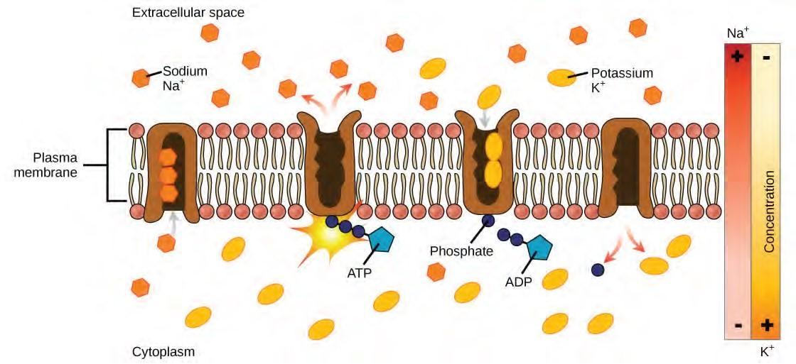
Figure 5.18 Primary active transport moves ions across a membrane, creating an electrochemical gradient (electrogenic transport). (credit: modification of work by Mariana Ruiz Villareal)
One of the most important pumps in animal cells is the sodium-potassium pump (Na+-K+ ATPase), which maintains the electrochemical gradient (and the correct concentrations of Na+ and K+) in living cells. The sodium-potassium pump moves K+ into the cell while moving Na+ out at the same time, at a ratio of three Na+ for every two K+ ions moved in. The Na+-K+ ATPase exists in two forms, depending on its orientation to the interior or exterior of the cell and its affinity for either sodium or potassium ions. The process consists of the following six steps.
- With the enzyme oriented towards the interior of the cell, the carrier has a high affinity for sodium ions. Three ions bind to the protein.
- ATP is hydrolyzed by the protein carrier and a low-energy phosphate group attaches to it.
- As a result, the carrier changes shape and re-orients itself towards the exterior of the membrane. The protein’s affinity for sodium decreases and the three sodium ions leave the carrier.
- The shape change increases the carrier’s affinity for potassium ions, and two such ions attach to the protein. Subsequently, the low-energy phosphate group detaches from the carrier.
- With the phosphate group removed and potassium ions attached, the carrier protein repositions itself towards the interior of the cell.
- The carrier protein, in its new configuration, has a decreased affinity for potassium, and the two ions are released into the cytoplasm. The protein now has a higher affinity for sodium ions, and the process starts again.
Several things have happened as a result of this process. At this point, there are more sodium ions outside of the cell than inside and more potassium ions inside than out. For every three ions of sodium that move out, two ions of potassium move in. This results in the interior being slightly more negative relative to the exterior. This difference in charge is important in creating the conditions necessary for the secondary process. The sodium-potassium pump is, therefore, an electrogenic pump (a pump that creates a charge imbalance), creating an electrical imbalance across the membrane and contributing to the membrane potential.
Visit the site (http://openstaxcollege.org/l/Na_K_ATPase) to see a simulation of active transport in a sodium-potassium ATPase.
Secondary Active Transport (Co-transport)
Secondary active transport brings sodium ions, and possibly other compounds, into the cell. As sodium ion concentrations build outside of the plasma membrane because of the action of the primary active transport process, an electrochemical gradient is created. If a channel protein exists and is open, the sodium ions will be pulled through the membrane. This movement is used to transport other substances that can attach themselves to the transport protein through the membrane (Figure 4.50). Many amino acids, as well as glucose, enter a cell this way. This secondary process is also used to store high-energy hydrogen ions in the mitochondria of plant and animal cells for the production of ATP. The potential energy that accumulates in the stored hydrogen ions is translated into kinetic energy as the ions surge through the channel protein ATP synthase, and that energy is used to convert ADP into ATP.
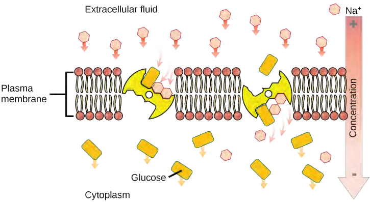
4.10 | Bulk Transport
In addition to moving small ions and molecules through the membrane, cells also need to remove and take in larger molecules and particles (see Table 4.3 for examples). Some cells are even capable of engulfing entire unicellular microorganisms. You might have correctly hypothesized that the uptake and release of large particles by the cell requires energy. A large particle, however, cannot pass through the membrane, even with energy supplied by the cell.
Endocytosis
Endocytosis is a type of active transport that moves particles, such as large molecules, parts of cells, and even whole cells, into a cell. There are different variations of endocytosis, but all share a common characteristic: The plasma membrane of the cell invaginates, forming a pocket around the target particle. The pocket pinches off, resulting in the particle being contained in a newly created intracellular vesicle formed from the plasma membrane.
Phagocytosis
Phagocytosis (the condition of “cell eating”) is the process by which large particles, such as cells or relatively large particles, are taken in by a cell. For example, when microorganisms invade the human body, a type of white blood cell called a neutrophil will remove the invaders through this process, surrounding and engulfing the microorganism, which is then destroyed by the neutrophil (Figure 4.51).
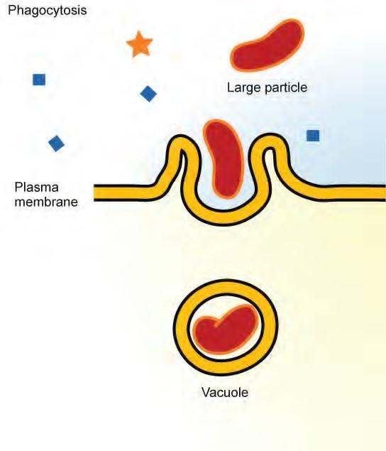
Figure 4.51 In phagocytosis, the cell membrane surrounds the particle and engulfs it. (credit:Mariana Ruiz Villareal)
In preparation for phagocytosis, a portion of the inward-facing surface of the plasma membrane becomes coated with a protein called clathrin, which stabilizes this section of the membrane. The coated portion of the membrane then extends from the body of the cell and surrounds the particle, eventually enclosing it. Once the vesicle containing the particle is enclosed within the cell, the clathrin disengages from the membrane and the vesicle merges with a lysosome for the breakdown of the material in the newly formed compartment (endosome). When accessible nutrients from the degradation of the vesicular contents have been extracted, the newly formed endosome merges with the plasma membrane and releases its contents into the extracellular fluid. The endosomal membrane again becomes part of the plasma membrane.
Pinocytosis
A variation of endocytosis is called pinocytosis. This literally means “cell drinking” and was named at a time when the assumption was that the cell was purposefully taking in extracellular fluid. In reality, this is a process that takes in molecules, including water, which the cell needs from the extracellular fluid. Pinocytosis results in a much smaller vesicle than does phagocytosis, and the vesicle does not need to merge with a lysosome (Figure 4.53).
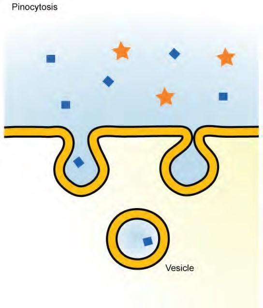
Figure 4.53 In pinocytosis, the cell membrane invaginates, surrounds a small volume of fluid, andpinches off. (credit: Mariana Ruiz Villareal)
A variation of pinocytosis is called potocytosis. This process uses a coating protein, called caveolin, on the cytoplasmic side of the plasma membrane, which performs a similar function to clathrin. The cavities in the plasma membrane that form the vacuoles have membrane receptors and lipid rafts in addition to caveolin. The vacuoles or vesicles formed in caveolae (singular caveola) are smaller than those in pinocytosis. Potocytosis is used to bring small molecules into the cell and to transport these molecules through the cell for their release on the other side of the cell, a process called transcytosis.
Receptor-mediated Endocytosis
A targeted variation of endocytosis employs receptor proteins in the plasma membrane that have a specific binding affinity for certain substances (Figure 4.53).
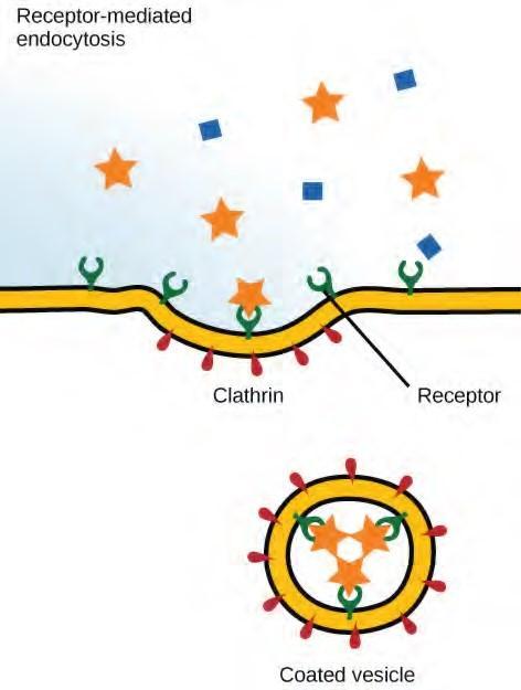
Figure 4.53 In receptor-mediated endocytosis, uptake of substances by the cell is targeted to a single type of substance that binds to the receptor on the external surface of the cell membrane.(credit: modification of work by Mariana Ruiz Villareal)
Although receptor-mediated endocytosis is designed to bring specific substances that are normally found in the extracellular fluid into the cell, other substances may gain entry into the cell at the same site. Flu viruses, diphtheria, and cholera toxin all have sites that cross-react with normal receptor-binding sites and gain entry into cells.
See receptor-mediated endocytosis in action, and click on different parts (http://openstaxcollege.org/ l/endocytosis) for a focused animation.
Exocytosis
The reverse process of moving material into a cell is the process of exocytosis. Exocytosis is the opposite of the processes discussed above in that its purpose is to expel material from the cell into the extracellular fluid. Waste material is enveloped in a membrane and fuses with the interior of the plasma membrane. This fusion opens the membranous envelope on the exterior of the cell, and the waste material is expelled into the extracellular space (Figure 5.23). Other examples of cells releasing molecules via exocytosis include the secretion of proteins of the extracellular matrix and secretion of neurotransmitters into the synaptic cleft by synaptic vesicles.
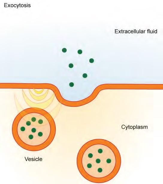
Figure 4.54 In exocytosis, vesicles containing substances fuse with the plasma membrane. The contents are then released to the exterior of the cell. (credit: modification of work by Mariana RuizVillareal)
Methods of Transport, Energy Requirements, and Types of Material
Transported
|
Transport Method |
Active/ Passive |
Material Transported |
|
Diffusion |
Passive |
Small-molecular weight material |
|
Osmosis |
Passive |
Water |
|
Facilitated transport/ diffusion |
Passive |
Sodium, potassium, calcium, glucose |
|
Primary active transport |
Active |
Sodium, potassium, calcium |
|
Secondary active transport |
Active |
Amino acids, lactose |
|
Phagocytosis |
Active |
Large macromolecules, whole cells, or cellular structures |
|
Pinocytosis and potocytosis |
Active |
Small molecules (liquids/water) |
|
Receptor-mediated endocytosis |
Active |
Large quantities of macromolecules |
Table 4.3
KEY TERMS
active transport method of transporting material that requires energy
amphiphilic molecule possessing a polar or charged area and a nonpolar or uncharged area capable of interacting with both hydrophilic and hydrophobic environments
antiporter transporter that carries two ions or small molecules in different directions
aquaporin channel protein that allows water through the membrane at a very high rate
carrier protein membrane protein that moves a substance across the plasma membrane by changing its own shape
caveolin protein that coats the cytoplasmic side of the plasma membrane and participates in the process of liquid update by potocytosis
channel protein membrane protein that allows a substance to pass through its hollow core across the plasma membrane
clathrin protein that coats the inward-facing surface of the plasma membrane and assists in the formation of specialized structures, like coated pits, for phagocytosis
concentration gradient area of high concentration adjacent to an area of low concentration
diffusion passive process of transport of low-molecular weight material according to its concentration gradient
electrochemical gradient gradient produced by the combined forces of an electrical gradient and a chemical gradient
electrogenic pump pump that creates a charge imbalance
endocytosis type of active transport that moves substances, including fluids and particles, into a cell
exocytosis process of passing bulk material out of a cell
facilitated transport process by which material moves down a concentration gradient (from high to low concentration) using integral membrane proteins
fluid mosaic model describes the structure of the plasma membrane as a mosaic of components including phospholipids, cholesterol, proteins, glycoproteins, and glycolipids (sugar chains attached to proteins or lipids, respectively), resulting in a fluid character (fluidity)
glycolipid combination of carbohydrates and lipids
glycoprotein combination of carbohydrates and proteins
hydrophilic molecule with the ability to bond with water; “water-loving” hydrophobic molecule that does not have the ability to bond with water; “water-hating”
hypertonic situation in which extracellular fluid has a higher osmolarity than the fluid inside the cell, resulting in water moving out of the cell
hypotonic situation in which extracellular fluid has a lower osmolarity than the fluid inside the cell, resulting in water moving into the cell
integral protein protein integrated into the membrane structure that interacts extensively with the hydrocarbon chains of membrane lipids and often spans the membrane; these proteins can be removed only by the disruption of the membrane by detergents
isotonic situation in which the extracellular fluid has the same osmolarity as the fluid inside the cell, resulting in no net movement of water into or out of the cell
osmolarity total amount of substances dissolved in a specific amount of solution
osmosis transport of water through a semipermeable membrane according to the concentration gradient of water across the membrane that results from the presence of solute that cannot pass through the membrane
passive transport method of transporting material through a membrane that does not require energy
peripheral protein protein found at the surface of a plasma membrane either on its exterior or interior side; these proteins can be removed (washed off of the membrane) by a high-salt wash
pinocytosis a variation of endocytosis that imports macromolecules that the cell needs from theextracellular fluid
plasmolysis detaching of the cell membrane from the cell wall and constriction of the cell membrane when a plant cell is in a hypertonic solution
potocytosis variation of pinocytosis that uses a different coating protein (caveolin) on the cytoplasmic side of the plasma membrane
primary active transport active transport that moves ions or small molecules across a membrane and may create a difference in charge across that membrane
pump active transport mechanism that works against electrochemical gradients
receptor-mediated endocytosis variation of endocytosis that involves the use of specific binding proteins in the plasma membrane for specific molecules or particles, and clathrin-coated pits that become clathrin-coated vesicles
secondary active transport movement of material that is due to the electrochemical gradient established by primary active transport
selectively permeable characteristic of a membrane that allows some substances through but not others solute substance dissolved in a liquid to form a solution
symporter transporter that carries two different ions or small molecules, both in the same direction
tonicity amount of solute in a solution
transport protein membrane protein that facilitates passage of a substance across a membrane by binding it
transporter specific carrier proteins or pumps that facilitate movement
uniporter transporter that carries one specific ion or molecule
cell theory see unified cell theory
cell wall rigid cell covering made of cellulose that protects the cell, provides structural support, and gives shape to the cell
central vacuole large plant cell organelle that regulates the cell’s storage compartment, holds water, and plays a significant role in cell growth as the site of macromolecule degradation
centrosome region in animal cells made of two centrioles
chlorophyll green pigment that captures the light energy that drives the light reactions of photosynthesis chloroplast plant cell organelle that carries out photosynthesis
chromatin protein-DNA complex that serves as the building material of chromosomes
chromosome structure within the nucleus that is made up of chromatin that contains DNA, the hereditary material
cilium (plural = cilia) short, hair-like structure that extends from the plasma membrane in large numbers and is used to move an entire cell or move substances along the outer surface of the cell
cytoplasm entire region between the plasma membrane and the nuclear envelope, consisting of organelles suspended in the gel-like cytosol, the cytoskeleton, and various chemicals
cytoskeleton network of protein fibers that collectively maintain the shape of the cell, secure some organelles in specific positions, allow cytoplasm and vesicles to move within the cell, and enable unicellular organisms to move independently
cytosol gel-like material of the cytoplasm in which cell structures are suspended
desmosome linkages between adjacent epithelial cells that form when cadherins in the plasma membrane attach to intermediate filaments
electron microscope an instrument that magnifies an object using a beam of electrons passed and bent through a lens system to visualize a specimen
endomembrane system group of organelles and membranes in eukaryotic cells that work together modifying, packaging, and transporting lipids and proteins
endoplasmic reticulum (ER) series of interconnected membranous structures within eukaryotic cells that collectively modify proteins and synthesize lipids
eukaryotic cell cell that has a membrane-bound nucleus and several other membrane-bound compartments or sacs
extracellular matrix material (primarily collagen, glycoproteins, and proteoglycans) secreted from animal cells that provides mechanical protection and anchoring for the cells in the tissue
flagellum (plural = flagella) long, hair-like structure that extends from the plasma membrane and is used to move the cell
gap junction channel between two adjacent animal cells that allows ions, nutrients, and low molecular weight substances to pass between cells, enabling the cells to communicate
Golgi apparatus eukaryotic organelle made up of a series of stacked membranes that sorts, tags, and packages lipids and proteins for distribution
intermediate filament cytoskeletal component, composed of several intertwined strands of fibrous protein, that bears tension, supports cell-cell junctions, and anchors cells to extracellular structures
light microscope an instrument that magnifies an object using a beam visible light passed and bent through a lens system to visualize a specimen
lysosome organelle in an animal cell that functions as the cell’s digestive component; it breaks down proteins, polysaccharides, lipids, nucleic acids, and even worn-out organelles
microfilament narrowest element of the cytoskeleton system; it provides rigidity and shape to the cell and enables cellular movements
microscope an instrument that magnifies an object
microtubule widest element of the cytoskeleton system; it helps the cell resist compression, provides a track along which vesicles move through the cell, pulls replicated chromosomes to opposite ends of a dividing cell, and is the structural element of centrioles, flagella, and cilia
mitochondria (singular = mitochondrion) cellular organelles responsible for carrying out cellular respiration, resulting in the production of ATP, the cell’s main energy-carrying molecule
nuclear envelope double-membrane structure that constitutes the outermost portion of the nucleus
nucleoid central part of a prokaryotic cell in which the chromosome is found
nucleolus darkly staining body within the nucleus that is responsible for assembling the subunits of the ribosomes
nucleoplasm semi-solid fluid inside the nucleus that contains the chromatin and nucleolus
nucleus cell organelle that houses the cell’s DNA and directs the synthesis of ribosomes and proteins
organelle compartment or sac within a cell
peroxisome small, round organelle that contains hydrogen peroxide, oxidizes fatty acids and amino acids, and detoxifies many poisons
plasma membrane phospholipid bilayer with embedded (integral) or attached (peripheral) proteins, and separates the internal content of the cell from its surrounding environment
plasmodesma (plural = plasmodesmata) channel that passes between the cell walls of adjacent plant cells, connects their cytoplasm, and allows materials to be transported from cell to cell
prokaryote unicellular organism that lacks a nucleus or any other membrane-bound organelle
ribosome cellular structure that carries out protein synthesis
rough endoplasmic reticulum (RER) region of the endoplasmic reticulum that is studded with ribosomes and engages in protein modification and phospholipid synthesis
smooth endoplasmic reticulum (SER) region of the endoplasmic reticulum that has few or no ribosomes on its cytoplasmic surface and synthesizes carbohydrates, lipids, and steroid hormones; detoxifies certain chemicals (like pesticides, preservatives, medications, and environmental pollutants), and stores calcium ions
tight junction firm seal between two adjacent animal cells created by protein adherence
unified cell theory a biological concept that states that all organisms are composed of one or more cells; the cell is the basic unit of life; and new cells arise from existing cells
vacuole membrane-bound sac, somewhat larger than a vesicle, which functions in cellular storage and transport
vesicle small, membrane-bound sac that functions in cellular storage and transport; its membrane is capable of fusing with the plasma membrane and the membranes of the endoplasmic reticulum and Golgi apparatus
CHAPTER SUMMARY
4.1 Studying Cells
A cell is the smallest unit of life. Most cells are so tiny that they cannot be seen with the naked eye. Therefore, scientists use microscopes to study cells. Electron microscopes provide higher magnification, higher resolution, and more detail than light microscopes. The unified cell theory states that all organisms are composed of one or more cells, the cell is the basic unit of life, and new cells arise from existing cells.
4.2 Prokaryotic Cells
Prokaryotes are predominantly single-celled organisms of the domains Bacteria and Archaea. All prokaryotes have plasma membranes, cytoplasm, ribosomes, and DNA that is not membrane-bound. Most have peptidoglycan cell walls and many have polysaccharide capsules. Prokaryotic cells range in diameter from 0.1 to 5.0 μm.
As a cell increases in size, its surface area-to-volume ratio decreases. If the cell grows too large, the plasma membrane will not have sufficient surface area to support the rate of diffusion required for the increased volume.
4.3 Eukaryotic Cells
Like a prokaryotic cell, a eukaryotic cell has a plasma membrane, cytoplasm, and ribosomes, but a eukaryotic cell is typically larger than a prokaryotic cell, has a true nucleus (meaning its DNA is surrounded by a membrane), and has other membrane-bound organelles that allow for compartmentalization of functions. The plasma membrane is a phospholipid bilayer embedded with proteins. The nucleus’s nucleolus is the site of ribosome assembly. Ribosomes are either found in the cytoplasm or attached to the cytoplasmic side of the plasma membrane or endoplasmic reticulum. They perform protein synthesis. Mitochondria participate in cellular respiration; they are responsible for the majority of ATP produced in the cell. Peroxisomes hydrolyze fatty acids, amino acids, and some toxins. Vesicles and vacuoles are storage and transport compartments. In plant cells, vacuoles also help break down macromolecules.
Animal cells also have a centrosome and lysosomes. The centrosome has two bodies perpendicular to each other, the centrioles, and has an unknown purpose in cell division. Lysosomes are the digestive organelles of animal cells.
Plant cells and plant-like cells each have a cell wall, chloroplasts, and a central vacuole. The plant cell wall, whose primary component is cellulose, protects the cell, provides structural support, and gives shape to the cell. Photosynthesis takes place in chloroplasts. The central vacuole can expand without having to produce more cytoplasm.
4.4 The Endomembrane System and Proteins
The endomembrane system includes the nuclear envelope, lysosomes, vesicles, the ER, and Golgi apparatus, as well as the plasma membrane. These cellular components work together to modify, package, tag, and transport proteins and lipids that form the membranes.
The RER modifies proteins and synthesizes phospholipids used in cell membranes. The SER synthesizes carbohydrates, lipids, and steroid hormones; engages in the detoxification of medications and poisons; and stores calcium ions. Sorting, tagging, packaging, and distribution of lipids and proteins take place in the Golgi apparatus. Lysosomes are created by the budding of the membranes of the RER and Golgi. Lysosomes digest macromolecules, recycle worn-out organelles, and destroy pathogens.
4.5 The Cytoskeleton
The cytoskeleton has three different types of protein elements. From narrowest to widest, they are the microfilaments (actin filaments), intermediate filaments, and microtubules. Microfilaments are often associated with myosin. They provide rigidity and shape to the cell and facilitate cellular movements. Intermediate filaments bear tension and anchor the nucleus and other organelles in place. Microtubules help the cell resist compression, serve as tracks for motor proteins that move vesicles through the cell, and pull replicated chromosomes to opposite ends of a dividing cell. They are also the structural element of centrioles, flagella, and cilia.
4.6 Connections between Cells and Cellular Activities
Animal cells communicate via their extracellular matrices and are connected to each other via tight junctions, desmosomes, and gap junctions. Plant cells are connected and communicate with each other via plasmodesmata.
When protein receptors on the surface of the plasma membrane of an animal cell bind to a substance in the extracellular matrix, a chain of reactions begins that changes activities taking place within the cell. Plasmodesmata are channels between adjacent plant cells, while gap junctions are channels between adjacent animal cells. However, their structures are quite different. A tight junction is a watertight seal between two adjacent cells, while a desmosome acts like a spot weld.
4.7 Components and Structure
The modern understanding of the plasma membrane is referred to as the fluid mosaic model. The plasma membrane is composed of a bilayer of phospholipids, with their hydrophobic, fatty acid tails in contact with each other. The landscape of the membrane is studded with proteins, some of which span the membrane. Some of these proteins serve to transport materials into or out of the cell. Carbohydrates are attached to some of the proteins and lipids on the outward-facing surface of the membrane, forming complexes that function to identify the cell to other cells. The fluid nature of the membrane is due to temperature, the configuration of the fatty acid tails (some kinked by double bonds), the presence of cholesterol embedded in the membrane, and the mosaic nature of the proteins and protein-carbohydrate combinations, which are not firmly fixed in place. Plasma membranes enclose and define the borders of cells, but rather than being a static bag, they are dynamic and constantly in flux.
4.8 Passive Transport
The passive forms of transport, diffusion and osmosis, move materials of small molecular weight across membranes. Substances diffuse from areas of high concentration to areas of lower concentration, and this process continues until the substance is evenly distributed in a system. In solutions containing more than one substance, each type of molecule diffuses according to its own concentration gradient, independent of the diffusion of other substances. Many factors can affect the rate of diffusion, including concentration gradient, size of the particles that are diffusing, temperature of the system, and so on.
In living systems, diffusion of substances into and out of cells is mediated by the plasma membrane. Some materials diffuse readily through the membrane, but others are hindered, and their passage is made possible by specialized proteins, such as channels and transporters. The chemistry of living things occurs in aqueous solutions, and balancing the concentrations of those solutions is an ongoing problem. In living systems, diffusion of some substances would be slow or difficult without membrane proteins that facilitate transport.
4.9 Active Transport
The combined gradient that affects an ion includes its concentration gradient and its electrical gradient. A positive ion, for example, might tend to diffuse into a new area, down its concentration gradient, but if it is diffusing into an area of net positive charge, its diffusion will be hampered by its electrical gradient. When dealing with ions in aqueous solutions, a combination of the electrochemical and concentration gradients, rather than just the concentration gradient alone, must be considered. Living cells need certain substances that exist inside the cell in concentrations greater than they exist in the extracellular space. Moving substances up their electrochemical gradients requires energy from the cell. Active transport uses energy stored in ATP to fuel this transport. Active transport of small molecular sized materials uses integral proteins in the cell membrane to move the materials: These proteins are analogous to pumps. Some pumps, which carry out primary active transport, couple directly with ATP to drive their action. In co-transport (or secondary active transport), energy from primary transport can be used to move another substance into the cell and up its concentration gradient.
4.10 Bulk Transport
Active transport methods require the direct use of ATP to fuel the transport. Large particles, such as macromolecules, parts of cells, or whole cells, can be engulfed by other cells in a process called phagocytosis. In phagocytosis, a portion of the membrane invaginates and flows around the particle, eventually pinching off and leaving the particle entirely enclosed by an envelope of plasma membrane.
Vesicle contents are broken down by the cell, with the particles either used as food or dispatched. Pinocytosis is a similar process on a smaller scale. The plasma membrane invaginates and pinches off, producing a small envelope of fluid from outside the cell. Pinocytosis imports substances that the cell needs from the extracellular fluid. The cell expels waste in a similar but reverse manner: it pushes a membranous vacuole to the plasma membrane, allowing the vacuole to fuse with the membrane and incorporate itself into the membrane structure, releasing its contents to the exterior.
ART CONNECTION QUESTIONS
Figure 4.7 Prokaryotic cells are much smaller than eukaryotic cells. What advantages might small cell size confer on a cell? What advantages might large cell size have?
Figure 4.8 If the nucleolus were not able to carry out its function, what other cellular organelles would be affected?
Figure 4.18 If a peripheral membrane protein were synthesized in the lumen (inside) of the ER, would it end up on the inside or outside of the plasma membrane?
REVIEW QUESTIONS
When viewing a specimen through a light microscope, scientists use ________ to distinguish the individual components of cells.
a. a beam of electrons
b. radioactive isotopes
c. special stains
d. high temperatures
The ________ is the basic unit of life.
a. organism
b. cell
c. tissue
d. organ
Prokaryotes depend on ________ to obtain some materials and to get rid of wastes.
a. ribosomes
b. flagella
c. cell division
d. diffusion
Which of the following is surrounded by two phospholipid bilayers?
a. the ribosomes
b. the vesicles
c. the cytoplasm
d. the nucleoplasm
Peroxisomes got their name because hydrogen peroxide is:
a. used in their detoxification reactions
b. produced during their oxidation reactions
c. incorporated into their membranes
d. a cofactor for the organelles’ enzymes
In plant cells, the function of the lysosomes is carried out by __________.
a. vacuoles
b. peroxisomes
c. ribosomes
d. nuclei
Which of the following is found both in eukaryotic and prokaryotic cells?
a. nucleus
b. mitochondrion
c. vacuole
d. ribosomes
Which of the following is not a component of the endomembrane system?
a. mitochondrion
b. Golgi apparatus
c. endoplasmic reticulum
d. lysosome
The process by which a cell engulfs a foreign particle is known as:
a. endosymbiosis
b. phagocytosis
c. hydrolysis
d. membrane synthesis
Which of the following is most likely to have the greatest concentration of smooth endoplasmic reticulum?
a. a cell that secretes enzymes
b. a cell that destroys pathogens
c. a cell that makes steroid hormones
d. a cell that engages in photosynthesis
Which of the following sequences correctly lists in order the steps involved in the incorporation of a proteinaceous molecule within a cell?
a. synthesis of the protein on the ribosome; modification in the Golgi apparatus; packaging in the endoplasmic reticulum; tagging in the vesicle
b. synthesis of the protein on the lysosome; tagging in the Golgi; packaging in the vesicle; distribution in the endoplasmic reticulum
c. synthesis of the protein on the ribosome; modification in the endoplasmic reticulum; tagging in the Golgi; distribution via the vesicle
d. synthesis of the protein on the lysosome; packaging in the vesicle; distribution via the Golgi; tagging in the endoplasmic reticulum
Which of the following have the ability to disassemble and reform quickly?
a. microfilaments and intermediate filaments
b. microfilaments and microtubules
c. intermediate filaments and microtubules
d. only intermediate filaments
Which of the following do not play a role in intracellular movement?
a. microfilaments and intermediate filaments
b. microfilaments and microtubules
c. intermediate filaments and microtubules
d. only intermediate filaments
Which of the following are found only in plant cells?
a. gap junctions
b. desmosomes
c. plasmodesmata
d. tight junctions
The key components of desmosomes are cadherins and __________.
a. actin
b. microfilaments
c. intermediate filaments
d. microtubules
Which characteristic of a phospholipid contributes to the fluidity of the membrane?
its head
cholesterol
a saturated fatty acid tail
double bonds in the fatty acid tail
What is the primary function of carbohydrates attached to the exterior of cell membranes?
identification of the cell
flexibility of the membrane
strengthening the membrane
channels through membrane
Water moves via osmosis _________.
throughout the cytoplasm
from an area with a high concentration of other solutes to a lower one
from an area with a high concentration of water to one of lower concentration
from an area with a low concentration of water to one of higher concentration
The principal force driving movement in diffusion is the __________.
temperature
particle size
concentration gradient
membrane surface area
What problem is faced by organisms that live in fresh water?
Their bodies tend to take in too much water.
They have no way of controlling their tonicity.
Only salt water poses problems for animals that live in it.
Their bodies tend to lose too much water to their environment.
Active transport must function continuously because __________.
plasma membranes wear out
not all membranes are amphiphilic
facilitated transport opposes active
transport
diffusion is constantly moving solutes in opposite directions
How does the sodium-potassium pump make the interior of the cell negatively charged?
by expelling anions
by pulling in anions
by expelling more cations than are taken in
by taking in and expelling an equal number of cations
What is the combination of an electrical gradient and a concentration gradient called?
potential gradient
electrical potential
concentration potential
electrochemical gradient
What happens to the membrane of a vesicle after exocytosis?
It leaves the cell.
It is disassembled by the cell.
It fuses with and becomes part of the plasma membrane.
It is used again in another exocytosis event.
Which transport mechanism can bring whole cells into a cell?
pinocytosis
phagocytosis
facilitated transport
primary active transport
In what important way does receptor mediated endocytosis differ from phagocytosis?
It transports only small amounts of fluid.
It does not involve the pinching off of membrane.
It brings in only a specifically targeted substance.
It brings substances into the cell, while phagocytosis removes substances.
CRITICAL THINKING QUESTIONS
In your everyday life, you have probably noticed that certain instruments are ideal for certain situations. For example, you would use a spoon rather than a fork to eat soup because a spoon is shaped for scooping, while soup would slip between the tines of a fork. The use of ideal instruments also applies in science. In what situation(s) would the use of a light microscope be ideal, and why?
Antibiotics are medicines that are used to fight bacterial infections. These medicines kill prokaryotic cells without harming human cells. What part or parts of the bacterial cell do you think antibiotics target? Why?
Explain why not all microbes are harmful.
You already know that ribosomes are abundant in red blood cells. In what other cells of the body would you find them in great abundance? Why?
What are the structural and functional similarities and differences between mitochondria and chloroplasts?
In the context of cell biology, what do we mean by form follows function? What are at least two examples of this concept?
In your opinion, is the nuclear membrane part of the endomembrane system? Why or why not? Defend your answer.
What are the similarities and differences between the structures of centrioles and flagella?
How do cilia and flagella differ?
How does the structure of a plasmodesma differ from that of a gap junction?
Explain how the extracellular matrix function
Why is it advantageous for the cell membrane to be fluid in nature?
Why do phospholipids tend to spontaneously orient themselves into something resembling a membrane?
Discuss why the following affect the rate of diffusion: molecular size, temperature, solution density, and the distance that must be traveled.
Why does water move through a membrane?
Both of the regular intravenous solutions administered in medicine, normal saline and lactated Ringer’s solution, are isotonic. Why is this important?
Where does the cell get energy for active transport processes?
How does the sodium-potassium pump contribute to the net negative charge of the interior of the cell?
Why is it important that there are different types of proteins in plasma membranes for the transport of materials into and out of a cell?
Chapter is adapted from:
Biology 2e by Mary Ann Clark, Matthew Douglas, and Jung Choi, published by OpenStax, licensed under CC BY 4.0, click here to access for free.
