
7.1 PreLab Reading – Muscle Physiology
Skeletal Muscle Physiology
Skeletal muscles, which attach to bones via tendons, are essential for movement and structural support. They consist of numerous muscle fibers containing contractile proteins, actin, and myosin, responsible for muscle contraction. Skeletal muscles are under voluntary control and are stimulated by somatic motor neurons. A motor unit consists of a motor neuron and the muscle fiber it controls. When an action potential (AP) from the motor neuron reaches a muscle fiber, it triggers a muscle twitch. The force of a muscle contraction depends on the number of motor units activated (recruitment) and the force generated by individual muscle fibers, which can increase through the summation.
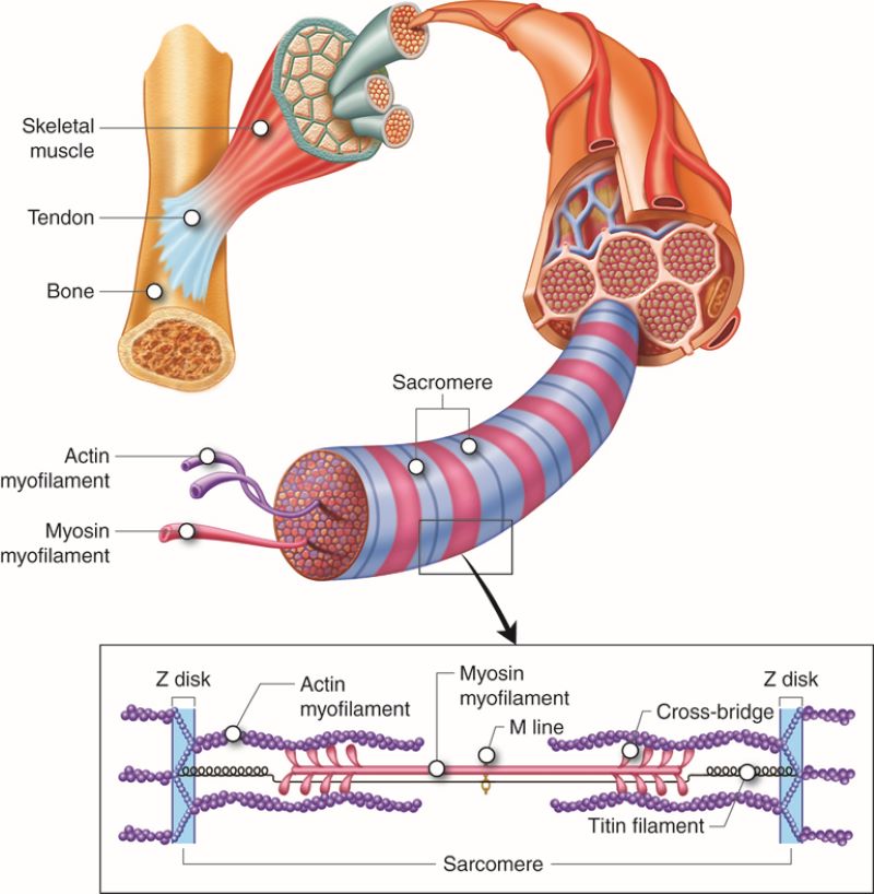
Diagram of a sarcomere showing the arrangement of actin (thin) and myosin (thick) filaments. Labels indicate the Z-lines, A-band, I-band, and the overlapping regions where contraction occurs. (Figure by https://pressbooks.ccconline.org/bio106/chapter/muscular-levels-of-organization/
During muscle contraction, multiple motor units fire repetitively. The electromyogram (EMG) records the electrical activity, which corresponds to the muscle contraction duration. The strength of the contraction is proportional to the electrical activity, which can be quantified by integrating the EMG spikes, creating an area under the curve that is linearly proportional to contraction strength.
Muscles can be stimulated through electrical impulses applied to motor points, where motor neurons enter the muscle. In this experiment, electrical stimulation will target the abductor digiti minimi (innervated by the ulnar nerve) and the flexor digitorum superficialis (innervated by the median nerve). These muscles play key roles in finger and wrist flexion. The motor points for these muscles are located on the medial surface of the palm, and lateral forearm (flexor digitorum superficialis). During the demonstration, your instructor will assist in locating these motor points to ensure accurate placement of the electrodes for effective stimulation.
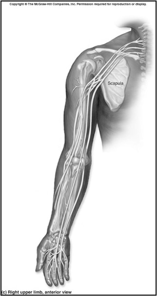
Diagram of the forearm showing motor points of two muscles: A flexor digitorium superficialis innervated by the median nerve, and the B- abductor digiti minimi innervated by the ulnar nerve. (Figure by McKinley, 1st edition).
In this week’s experiment, students will measure grip strength using a hand dynamometer and record EMG activity from forearm muscles. The relationship between EMG activity and grip strength will be analyzed by plotting maximum grip strength against the area under the EMG curve. Data will be collected from both forearms to compare muscle strength and electrical activity relative to forearm circumference. The rate of fatigue will also be assessed by recording prolonged grip strength and EMG activity.
Excitation-Contraction Coupling
Skeletal muscle fibers contain myofibrils, which consist of sarcomeres, the functional units of contraction. Each sarcomere is composed of actin (thin filaments) and myosin (thick filaments).
When a motor neuron stimulates a muscle fiber, it releases acetylcholine (Ach) from its terminal bouton at the motor end plate. ACh binds to Nicotinic Cholinergic receptors, triggering a graded depolarization known as the end-plate potential (EPP). The EPP is large enough to depolarized the membrane to threshold, generating an action potential (AP). The AP propagates along the sarcolemma and travels down into the muscle fiber through transverse tubules (T tubules). Within the T tubules, voltage-sensitive dihydropyridine (DHP) receptors detect the electrical signal and undergo a conformational change. This change mechanically interacts with ryanodine receptors on the sarcoplasmic reticulum, causing the release of calcium ions into the cytoplasm.
The released calcium binds to troponin on the acting filaments, shifting tropomyosin and exposing myosin binding sites. This initiates the Sliding Filament Theory, in which two sets of protein filaments, acting and myosin, slide past one another to shorten the sarcomere, causing the muscle fiber to contract. In striated muscle types such as skeletal and cardiac muscle, this sliding filament mechanism occurs through the Cross Bridge Cycle (CBC):
- Cross-Bridge Formation – Myosin heads bind to actin, forming a cross-bridge.
- Power Stroke – The splitting of ATP causes the myosin head to change shape and pull against actin.
- Cross-Bridge Detachment – A new ATP molecule binds to myosin, causing it to detach from actin.
- Myosin Reactivation – ATP is hydrolyzed, resetting myosin to its high-energy state for another cycle.
This repeated cycling of myosin-actin interactions shortens the sarcomere and produces muscle contraction.
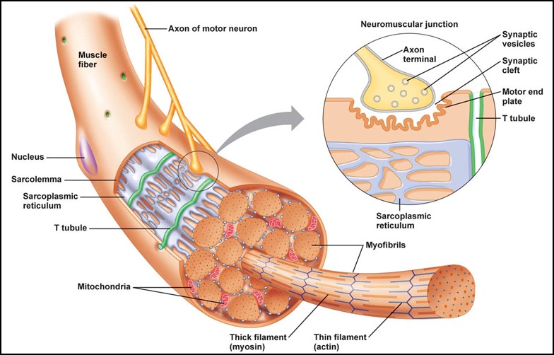
Diagram of a skeletal muscle fiber showing the neuromuscular junction, including the motor neuron terminal, synaptic cleft, and muscle fiber membrane, illustrating where nerve signals trigger muscle contraction. (Figure by Standfield, 4th edition).
The strength of the contraction depends on the amount of calcium released and the number of crossbridge formed. When muscles are stimulated repeatedly, twitches can combine into a sustained contraction, or tetanus, if the stimulation frequency is high enough that the muscle fibers don’t fully relax.
Muscle Twitch
A muscle twitch is the mechanical response of a muscle fiber to a single stimulus and consists of three distinct phases:
- Latent Phase – This initial period occurs between the AP in the muscle fiber and the onset of contraction. During this phase, the sarcolemma and T-tubules depolarize, triggering the conformational change in DHP receptors, which in turn activates Ryanodine receptors on the sarcoplasmic reticulum. This leads to Ca2+ release, allowing myosin to bind to actin, and the early development of tension.
- Contraction Phase – This phase begins when the muscle starts generating force and continues until it reaches peak tension. Calcium levels in the cytoplasm rise, facilitating cross-bridge cycling between action and myosin, which leads to sarcomere shortening and increased muscle tension.
- Relaxation Phase – The longest phase, during which Ca2+ reuptake into the sarcoplasmic reticulum exceeds its release. As intracellular calcium decrease, and muscle tension gradually diminishes until the fiber returns to its resting state.
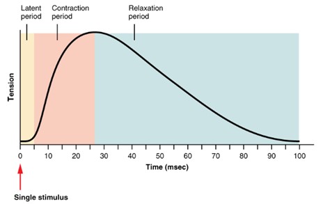
Graph showing the phases of a muscle Twitch: latent phase (time between stimulus and contraction), contraction phase (muscle tension increases), and relaxation phase (muscle tension decreases back to resting level). (Figure by OpenStax, used under a Creative Commons Attribution license.)
There are two types of muscles twitches:
- Isometric: The muscle generates tension but does not no change length (e.g., holding a plank).
- Isotonic: The muscle shortens as it lifts a load (e.g., bicep curls).
Isotonic contractions begin with an isometric phase where the muscle contracts without shortening until enough force is generated.
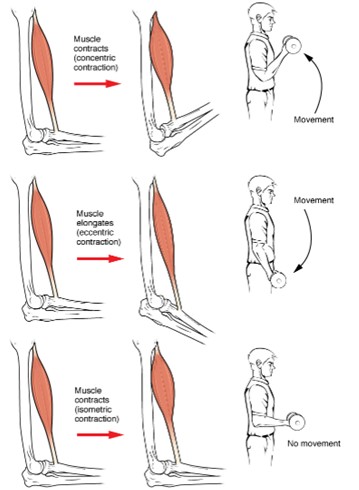
Diagram comparing isotonic and isometric muscle contractions. In isotonic contraction, muscle length changes while tension remains constant. In isometric contraction, muscle length stays the same while tension increases. (Figure by OpenStax, used under a Creative Commons Attribution license.)
Diagram comparing isotonic and isometric muscle contractions. In isotonic contraction, muscle length changes while tension remains constant. In isometric contraction, muscle length stays the same while tension increases.
Excitation-Contraction Coupling and Muscle Fatigue
Excitation-contraction coupling converts neural signals into muscle contraction. When a motor neuron releases acetylcholine, it triggers an action potential in the muscle fiber, leading to the processes outlined above.
Frequently stimulation leads to muscle fatigue, influenced by factors such as exercise intensity, the muscle fiber type, and blood supply. For example, high-intensity exercise recruits fast glycolytic fibers that generate ATP via glycolysis, leading to lactic acid buildup and fatigue. Whereas, prolonged low-intensity exercise depletes glycogen stores, leading to fatigue in slow-oxidative fibers.
ATP is required for both muscle contraction and relaxation. Without ATP, calcium ions remain in the cytoplasm, keeping actin and myosin bound, preventing relaxation.
Motor Unit Recruitment
Skeletal muscles are made of motor units, which consist of a motor neuron and the muscle fibers it innervates. Small motor units (with smaller somas) are recruited first, responding to weaker stimuli, while larger motor units (with larger somas) are recruited as the stimulus increases. The force of contraction depends on the number and size of motor units recruited.
Motor units are activated asynchronously to prevent fatigue. Recruitment is increased by the frequency of stimulation, a process known as summation. If a second stimulus is applied before the muscle has relaxed from the first, it leads to greater force production. If the frequency is high enough, the muscle may experience tetanus – either incomplete or complete – where the muscle maintains a steady contraction.
Fiber Types and Muscle Contraction
Muscle fibers are classified into three types: fast glycolytic, fast oxidative, and slow oxidative. Fast glycolytic fibers generate the most force but fatigue quickly. Fast oxidative fibers are most resistant to fatigue, and slow oxidative are the most enduring but generate the least force.
Muscles typically contain a mix of these fibers. When higher force is required, larger motor units with fast glycolytic fibers are recruited. The recruitment order follows slow oxidative first, fast oxidative second, and fast glycolytic last.
Effect of Load on Isotonic Contractions
In isotonic contractions, the muscle generates enough tension to overcome a load, causing it to shorten. As the load increases:
- The latent period (time before shortening) becomes longer.
- The duration of shortening decreases.
- The velocity of shortening slows down.
The muscle’s work output is affected by the load, as work is the product of force and distance. No work is done in an isometric contraction, where no distance is covered.
Summation and Tetanus
Summation occurs when successive stimuli increase muscle tension. This happens because calcium ions remain in the cytoplasm after a previous stimulus, allowing for more myosin-actin binding. With frequent stimulation, the muscle fiber reaches incomplete tetanus, where individual twitches are still observable, but tension is higher than a single twitch. With further stimulation, the muscle reaches complete tetanus, where no relaxation occurs, leading to maximum tension.
Complete tetanus results in sustained contraction, and relaxation occurs more slowly as calcium is pumped back into the sarcoplasmic reticulum. Under normal conditions, motor units fire asynchronously, allowing for smooth, sustained muscle contractions.
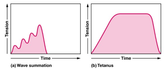
Graph showing muscle tension over time with repeated stimuli. Individual twitches summate to produce higher tension, leading to incomplete tetanus and, with rapid stimulation, complete tetanus where tension reaches a sustained maximum. (Figure by OpenStax, used under a Creative Commons Attribution license.)
Muscle Fatigue
Muscle fatigue, a decrease in tension production following frequent stimulation, is influenced by exercise type, contraction rate, force, and blood supply. High-intensity activities like weightlifting recruit fast glycolytic fibers, which depend on fermentation (glycolysis) for ATP. This process generates lactic acid, altering pH and disrupting the function of contractile proteins and calcium pumps in the sarcoplasmic reticulum. Intense exercise may also lead to neuromuscular fatigue, where somatic motor neurons deplete their neurotransmitter supply. In contrast, low-intensity, long duration exercises primarily fatigue slow oxidative and fast oxidative fibers through fuel source depletion, such as glycogen.
Energy is crucial not only for contraction but also for relaxation. Calcium ions must actively return to the sarcoplasmic reticulum, a process requiring ATP. Without adequate ATP, calcium remains in the cytoplasm, prolonging actin-myosin interaction and preventing efficient relaxation.
In this lab, students will explore excitation-contraction coupling by stimulating the motor points of the abductor digiti minimi and flexor digitorium superficialis to observe muscle contraction via EMG. Analyzing grip strength and EMG activity reveals the connection between electrical impulses and muscle force. Comparisons between dominant and non-dominant forearms provide insights into muscle size, electrical activity, and fatigue, highlighting the physiological basis of muscle performance.
Questions
-
- What term describes the connection between a somatic motor neuron and the muscle fibers it innervates?
- How is the strength of a muscle contraction related to the number of motor units recruited?
- Describe the following terms related to muscle physiology and their corresponding functions or characteristics:
- Muscle fibers
- Myofibrils
- Sarcomere
- T tubules
- Sarcoplasmic reticulum
- Latent period
- Isometric contraction
- Isotonic contraction
- Describe the three types of muscle fibers, including how they generate energy, their force production, and their susceptibility to fatigue.
- What is the term for the process in which a second muscle twitch adds to the first when a muscle fiber is stimulated before completing its current twitch?
Adapted from Human Physiology Lab Manual by Jim Blevins, Melaney Farr, and Arleen Sawitzke, Salt Lake Community College.
