
6.1 PreLab Reading – The Endocrine System
In this lab, we will build upon our previous knowledge to explore how cells communicate with target cells. Chemical messengers, also known as signaling molecules, can be classified based on their function or chemical structure:
There are 5 major classes of chemical messengers in the human body:
- Amino Acid Messengers: There are four main types: glutamine, aspartate, glycine, and gamma aminobutyric acid (GABA). These molecules are hydrophilic and must be synthesized within the neuron that secretes them. Because they are produced within neurons, they are referred to as neurotransmitters. They are synthesized in the cytoplasm and then packaged into vesicles for secretion.
- Amine Messengers: These messengers all contain an amino group. Some are catecholamines, which are derived from the amino acid tyrosine. Examples include dopamine and norepinephrine (both neurotransmitters), as well as epinephrine, which typically functions as a hormone. All of the amines, with the exception of T3/T4, are hydrophilic.
- Peptide/Protein Messengers: Proteins are polymers of amino acids. The distinction between peptides and proteins lies in their length: peptides consist of fewer than 50 amino acids bonded together while proteins are composed of 51 or more amino acids. This category encompasses a vast array of signaling molecules within the human body. Peptides and proteins are hydrophilic due to their amino acid composition. Examples include insulin (regulates blood glucose levels), TRH (stimulates the release of TSH), ACTH (stimulates the secretion of glucocorticoids such as cortisol), ADH (increases water reabsorption). All amino acids are hydrophilic and, therefore, peptide/proteins are hydrophilic.
- Steroid Messenger: Steroids are hydrophobic molecules typically composed of three 6-carbon rings and one 5-carbon ring. Derived from cholesterol and synthesized in the smooth endoplasmic reticulum, steroids function primarily as hormones. Due to their hydrophobic nature, steroids can diffuse across cell membranes but cannot be stored; they must be synthesized upon demand. Examples include testosterone, estrogen, cortisol, aldosterone, and Vitamin D.
- Eicosanoid Messengers: Eicosanoids are hydrophobic lipids, functioning as small signaling molecules that can pass through cell membranes. They cannot be stored and are synthesized as needed. Most eicosanoids are paracrine in nature and can be produced by nearly all cells in the body. They are primarily synthesized from arachidonic acid, a 20-carbon fatty acid derived from cell membrane phospholipids or second messengers like diacylglycerol (DAG). Eicosanoids follow two main pathways: the Cyclooxygenase (COX) pathway and the Lipoxygenase pathway. Examples include prostacyclin and thromboxane (involved in blood clotting), prostaglandins (involved in various systems including inflammation), and leukotrienes (promote inflammation). Certain anti-inflammatory drugs inhibit these synthesis pathways; for instance, aspirin inhibits cyclooxygenase (COX), a crucial enzyme in prostaglandin production.
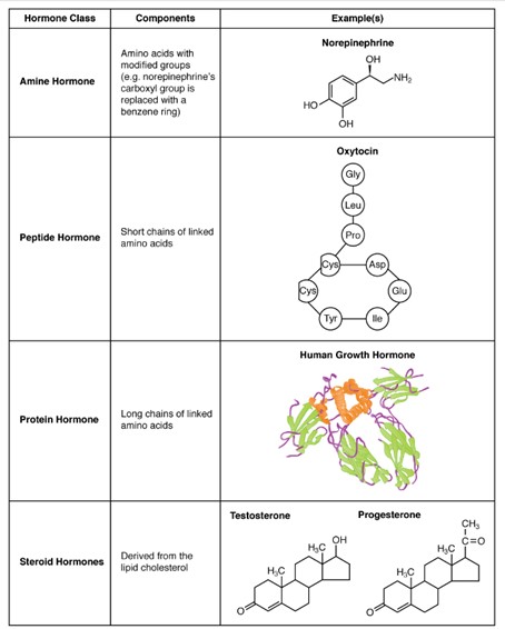
Chemical Signal Classes: Amine, peptide, protein, and steroid. Eicosanoids are not pictures (Figure by OpenStax is used under a Creative Commons Attribution license.)
Endocrine System
A hormone functions as a chemical messenger that travels from its source (secretory) cell to target cells via the bloodstream. Typically secreted by endocrine glands (such as the adrenals, thyroid, and pituitary), hormones can also be produced by various cell types throughout the body.
Upon secretion, hydrophilic messengers like peptides/proteins, and non-thyroid hormone amines dissolve directly into the watery blood plasma, circulating briefly before being metabolized by the liver and excreted by the kidneys, typically within minutes. In contrast, hydrophobic hormones such as thyroid hormones (T3/T4) and steroids require carrier proteins (such as corticosteroid-binding globulin and albumin) to travel through the bloodstream. Bound to these carriers, their half-lives are extended to hours or more.
The body’s organs coordinate activities through two primary systems: the nervous and endocrine systems. The nervous system uses rapid action potentials and neurotransmitters (paracrine or direct signaling) to convey information to and from the brain. Conversely, the endocrine system employs hormones, chemical messengers produced by specific tissues, which travel through the bloodstream to influence distant target organs.
Analogously, interpersonal communication can be compared to the difference between telephone calls and postal mail. The nervous system functions like a telephone, transmitting rapid, direct messages, while the endocrine system resembles the postal service, delivering messages more slowly. Although hormones travel throughout the body via the bloodstream, they only influence cells with specific receptors. Despite their slower transmission, hormonal effects can last for extended periods.
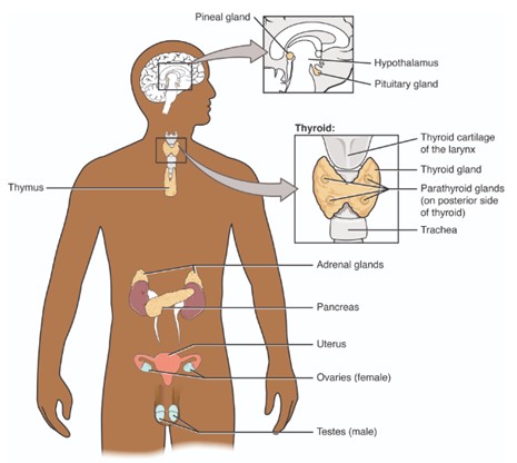
The Endocrine System: Primary organs of the endocrine system. (Figure by OpenStax is used under a Creative Commons Attribution license.)
The hypothalamus
The command center of the endocrine system is the hypothalamus, a small region about the size of a penny located in the brain. Composed of neural tissue rather than epithelial tissue, the hypothalamus integrates neural stimuli and releases neurohormones (hormones that is secreted by a neuron) to the endocrine system. These neurohormones travel through the bloodstream to their targets, exemplifying their dual role as neural and hormonal messengers.
The hypothalamus functions as an endocrine organ, secreting oxytocin and antidiuretic hormone (ADH; also known as vasopressin), both of which are peptides. These hormones are synthesized in hypothalamic cell bodies and transported down axons within the pituitary stalk (infundibulum) to their axon terminals in the posterior pituitary. From there, they are released directly into the bloodstream.
Furthermore, the hypothalamus regulates the function of the anterior pituitary gland by secreting releasing hormones such as thyrotropin-releasing hormone (TRH), corticotropin-releasing hormone (CRH), and gonadotropin-releasing hormone (GnRH). These releasing hormones travel through a specialized blood vessel system known as the hypothalamic to anterior pituitary (hypophyseal) portal system. Unlike the posterior pituitary, which consists of axon terminals from hypothalamic neurons and is not considered a gland, the anterior pituitary is glandular, composed of epithelial tissue.
In response to these releasing hormones, the anterior pituitary synthesizes and secretes various hormones including thyroid stimulating hormone (TSH), luteinizing hormone (LH), follicle stimulating hormone (FSH), growth hormone (GH), adrenocorticotropic hormone (ACTH), and prolactin. Each hormone enters the bloodstream to exert specific effects on target organs. For instance, TRH from the hypothalamus stimulates TSH release from the anterior pituitary, which then prompts the thyroid gland (the target organ) to release thyroid hormones (T3 and T4).
It’s important to note that hypothalamic releasing hormones are essential only for the synthesis and release of anterior pituitary hormones. In contrast, hormones stored and released by the posterior pituitary, such as oxytocin and vasopressin, are synthesized in the hypothalamus and transported along axons to the posterior for direct release into the bloodstream. Due to its role in secreting multiple hormones, the anterior pituitary gland is often referred to as the “master gland”.
Negative Feedback
Hormones release by the endocrine system are regulated through negative or positive feedback mechanisms. Negative feedback operates similarly to a thermostat set a predetermined temperature (e.g., 68° F). When the temperature exceeds the set point (e.g., 72° F), the thermostat activates the air conditioner to cool the room. Once the temperature drops below the set point (e.g., 67° F), the air conditioner turns off. This continuous assessment ensures the room remains at a relatively constant temperature.
In the endocrine system, feedback loops regulate hormone release by signaling the producing organ when sufficient levels have been reached, preventing excess production. Negative feedback can inhibit not only the releasing organ but also the pituitary gland (short loop) and hypothalamus (long loop), ensuring efficient hormone regulation while conserving resources.
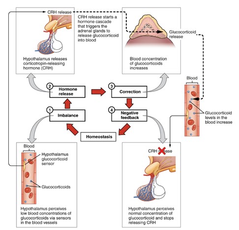
Negative Feedback: Hypothalamus responding to low blood concentration of glucocorticoids. (Figure by OpenStax is used under a Creative Commons Attribution license.)
Thyroid, Cortisol, and Testosterone
In this lab, we examine the pathways of three hormones: thyroid hormone, cortisol, and testosterone. The hormonal pathways are similar in all three cases. The hypothalamus plays a crucial role by secreting a releasing hormone to regulate each hormone secreted by the anterior pituitary gland. Think of the hypothalamus as a command center. Without stimulation, the hypothalamus will not secrete the release hormones (TRH, CRH, and GnRH) needed to stimulate the anterior pituitary gland.
The hypothalamus releases TRH, which travels via the bloodstream to the anterior pituitary gland, stimulating the production of TSH. TSH then travels to the thyroid gland (located anterior to the trachea and just below the larynx) to stimulate the production and release of thyroid hormones (T3 and T4). These hormones influence the growth rate of many body tissues and are necessary for proper central nervous system development. Their primary function is to increase basal metabolic rate (BMR) and heat production. Once released in the bloodstream, thyroid hormones provide feedback to the hypothalamus, anterior pituitary gland, and the thyroid gland by reducing the secretion of TRH, TSH, and thyroid hormones to maintain hormonal balance.
Similarly, ACTH is released from the anterior pituitary gland in response to CRH secreted by the hypothalamus. ACTH stimulates the adrenal cortex of the adrenal glands (located on top of the kidneys) to secrete cortisol, which promotes the breakdown of proteins and fats and helps the body adapt to stress. Cortisol provides the body with fuel by breaking down (catabolism) bodily materials. Plasma cortisol levels are typically regulated through negative feedback on the hypothalamus, anterior pituitary gland, and the adrenal gland by reducing the secretion of CRH, ACTH, and cortisol to maintain balance. The release of CRH is regulated by negative feedback, circadian rhythms, and stress. Cortisol can also act as an immunosuppressive and anti-inflammatory agent. If administered in large doses, its immunosuppressive properties will cause the organs of the immune system to shrink. In this lab, the thymus gland will represent the organ of the immune system.
LH is released from the anterior pituitary gland in response to GnRH (gonadotropin-releasing hormone) secreted by the hypothalamus. LH is secreted in both males and females. In males, LH travels to the Leydig cells of the testes, which releases testosterone, one of the three predominant sex hormones in males. Testosterone is responsible for the male sex drive and secondary sex characteristics, such as increased body hair and a deeper voice. An excess of testosterone can cause an increase (anabolic) in muscle mass. Negative effects of testosterone include male pattern baldness and increased secretion of the sebaceous glands, which can lead to acne.
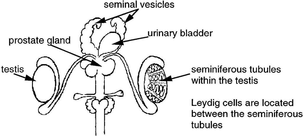
Organs of the Male Reproductive Tract : Prostate gland, testis, and seminiferous tubules. (Figure by OpenStax is used under a Creative Commons Attribution license.)
To simplify the relationship between the reproductive and endocrine systems, we will focus only on the male system. The female reproductive system is more challenging to study due to its continuous cycling.
The pathways of all three hormones can be understood by examining Figure 4, which also demonstrates the hormonal pathways used throughout the lab. This visual representation serves as an essential aid in analyzing laboratory data.
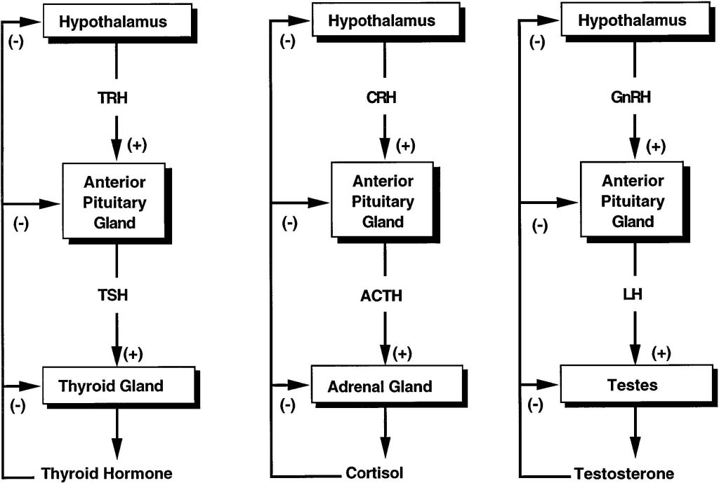
Figure 4: Negative feedback control (Hormone pathways)
The glands and tissues of our body enlarge (hypertrophy) when they are continuously activated. For example, a person who lifts weights regularly stimulates their muscles, resulting in muscle hypertrophy. This is easily observed when comparing a bodybuilder to an average person; the bodybuilder’s muscles are significantly larger. In contrast, if a gland or tissue is continuously inhibited, it will shrink or atrophy. For instance, if a cast is placed on a person’s arm for six weeks and then removed, a noticeable reduction in muscle mass will be seen. The lack of movement (stimulation) of the limb during this period leads to muscle atrophy.
Abnormal hormone secretion can have serious consequences. Some diseases result from excessive hormone production, known as hypersecretion, while others arise from insufficient hormone production, referred as hyposecretion. These imbalances can have significant effects on bodily functions and overall health.
Understanding these hormonal pathways and their imbalances is crucial for diagnosing and treating related diseases effectively.
Examples
Hyperthyroidism and Hypothyroidism
Hyperthyroidism is characterized by the excessive production of thyroid hormones. The most common cause is Graves’ disease. Symptoms of hyperthyroidism include:
- Increased basal metabolic rate (BMR)
- Feeling hot
- Nervousness
- Weight loss
- Enlarged thyroid gland (goiter)
Hypothyroidism result from decreased levels of thyroid hormones. Symptoms include:
- Low BMR
- Feeling cold
- Tiredness
- Weight gain
Cushing’s Syndrome and Addison’s Disease
Cushing’s syndrome is caused by the overproduction of cortisol (hypercortisolism). Symptoms include:
- Personality changes
- Hypertension (high blood pressure)
- Osteoporosis (bone weakening due to calcium loss)
- Initial weight loss, followed by long-term weight gain as metabolism drops
Excess cortisol leads to protein degradation, resulting in muscle mass loss and a “wasting” effect.
Addison’s disease is characterized by the underproduction of cortisol (hypocortisolism). Symptoms include:
- Defective metabolism
- Mental confusion
- Decreased ability to adapt to stress
Testosterone Imbalance
- Low testosterone levels primarily affect the sexual organs. In males, this can result in abnormal development and low sperm counts, impairing fertility.
- Excess testosterone is rate but can cause premature sexual development.
Questions
-
- Which type of feedback inhibits further hormone secretion?
- Which hormone plays the major role in increasing body-wide basal metabolic rate (BMR)?
- Which hormone acts as both an immunosuppressive and anti-inflammatory agent?
- What is muscle atrophy, and what are some common causes of it?
- What is the term for excessive production of thyroid hormones?
Adapted from Human Physiology Lab Manual by Jim Blevins, Melaney Farr, and Arleen Sawitzke, Salt Lake Community College.
