
5.1 PreLab Reading – The Sensory System
The Nervous System
The nervous system is divided into two main divisions: the central nervous system (CNS) and the peripheral nervous system (PNS). The CNS, consisting of the brain and spinal cord, serves as the body’s primary control center. The PNS, which includes all neurons outside the CNS, facilitates communication between the CNS and the rest of the body through afferent and efferent pathways.
The CNS integrates sensory input and generates appropriate motor responses. The PNS, in turn, is divided into afferent and efferent branches. The afferent branch carries sensory information (somatic, special, and visceral senses) to the CNS, while the efferent branch transmits motor commands from the CNS to effector organs.
The efferent branch is further divided into the somatic and autonomic nervous systems. The somatic nervous system controls voluntary movements by regulating skeletal muscle contractions. The autonomic nervous system (ANS) governs involuntary functions by regulating smooth muscle, cardiac muscle, adipose tissue, and glands. The ANS is further divided into the parasympathetic and sympathetic nervous systems, which work together to maintain homeostasis.
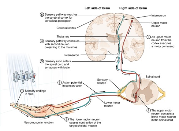
Sensory System: Sensory input received and transfer to the CNS for proper sensory perception. (Figure by OpenStax is used under a Creative Commons Attribution license.)
Neurons
We first introduced the basic structure of neurons in Lab 4. Here, we will review that foundation and explore additional details.
Neurons are the fundamental functional units of the nervous system. These excitable cells generate actions potentials, which are rapid, large electrical signals that travel long distances. Given their specialization, it is important to understand their key structural components:
- Cell body (soma): This part of the cell resembles a typical cell, containing the nucleus and organelles necessary for normal metabolic functions. The soma can receive inputs in the form of chemical, electrical, or non-chemical stimuli (such as light or sound). Interestingly, when most CNS neurons become fully mature (G0 stage), they lose the ability to divide because they shed their centrioles.
- Dendrites: These are small, branch-like extensions of the plasma membrane that typically branch off from the soma. Dendrites are specialized to receive inputs in the form of chemical stimulus or sensory stimulus.
- Axon (nerve fiber): These long strands, primarily composed of plasma membrane, are specialized for transmitting information. Most neurons have one main axon, but these can branch to transmit information to multiple sites. The portion of the axon that extends from the cell body is called the axon hillock and is specialized to initiate action potentials. The end of the axon, known as the axon terminal, is specialized to release neurotransmitters in response to action potentials traveling down the axon.
Sensory Systems and Sensory Pathways
Sensory receptors allow us to detect environmental stimuli and relay this information to the brain. The sensory systems include:
- Somatosensory system: Processes touch, temperature, pain, and limb position.
- Special senses: It includes vision, hearing, balance, taste, and smell.
In this lab, we will explore sensory systems that help us perceive the world around us. Sensory receptors in the skin, mouth, eyes, and nose detect stimuli and convert them into electrical and chemical signals that are sent to the brain. The brain then processes this information and generates appropriate responses.
A sensory pathway begins when a stimulus (light, touch, sound, or chemicals) alters the membrane potential (Vm) by opening of closing stimulus-gated ion channels in a receptor. The receptor, either a specialized neuron or a cell that communicates with an afferent neuron, generates an action potential that travels to the CNS. There, an interneuron relays the signal to the appropriate processing center.
Perception
Perception is how the brain interprets and categorizes sensory input. For example, the scent of a rose involves sensory receptors, neurons, and brain processing, which may lead to an action (e.g., sneezing or leaning in for a deeper sniff). However, perception is subjective. It can influenced by memory, emotions, and physiological state. The same scent might trigger pleasant nostalgia or, in a different circumstances, an unpleasant response.
Introduction to Our Lab
Our perception of reality is shaped by how we sense and interpret the world around us. These perceptions stem from sensory information processed by specialized receptors in the peripheral nervous system. In this lab, we will explore four of our primary senses: taste, touch, sight, and smell and how they contribute to our understanding of our surroundings.
Sensory receptors are specific for a particular type of stimuli including light (photoreceptors), chemicals (chemoreceptors), temperature changes (thermoreceptors), pressure (mechanoreceptors), and pain (nociceptors). When activated, these receptors convert stimuli into electrical signals through a process known as sensory transduction. The mechanism of transduction varies depending on the type of receptor.
Sensory signals travel along distinct labeled lines, which each type of sensory information following a specific neural pathway dedicated to that sensation. These labeled lines help the brain identify the nature of the sensory stimulus. For example, visual information follows a different pathway than olfactory (smell) signals. Once these sensory signals reach the brain via their respective labeled lines, they are processed and interpreted.
Most sensory signals is initially routed through the thalamus before being directed to specific regions of the cerebral cortex for further interpretation.
However, perception is not always an exact reflection of reality. Common phrase like “I can’t believe my eyes” or “My mind is playing tricks on me” highlight the brain’s potential to misinterpret sensory information. Through a series of activities, you will explore the limitations of your senses and how the brain processes sensory input.
Touch
The skin (integumentary system) is the body’s largest sensory organ, with numerous receptor sites for cutaneous sensations (form the Latin cutis, meaning “skin”). Different types of receptors in the skin respond to various stimuli, including:
- Mechanoreceptors: Detect touch and pressure
- Thermoreceptors: Sense temperature changes
- Nociceptors: Sense pain
The accuracy of tactile perception depends on which sensory receptors are activated and their pathways to the brain. Areas with high concentration of receptors can localize stimuli more precisely than areas within fewer receptors. This concept is demonstrated through the two-point discrimination test, which measures the smallest distance at which two separate stimuli can be distinguished as distinct. Body regions with a high density of mechanoreceptors, such as fingertips and lips, have finer two-point discrimination compared to areas like the back of the neck or mid-calf.
Once a stimulus is detected, nerve endings in the skin transmit the information to the spinal cord and then to the brain, where it is interpreted. Most somatic sensations are processed in the somatosensory cortex of the parietal lobe. The amount of cortical space dedicated to different body regions correlates with the sensitivity of those areas, meaning regions like the fingertips and lips have a higher concentration of receptors and more precise sensory perception than other parts of the body.
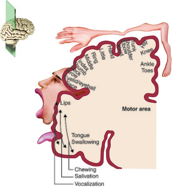
Somatosensory Cortex: Receptor densities and the corresponding size of receptive fields. (Figure by Wikipedia is used under a Creative Commons Attribution license.)
Vision
The eyes function as the primary sensory organ of the human visual system, allowing for the detection of light and the formation of detailed images. Light waves first pass through the cornea, aqueous humor, lens, and vitreous body before reaching the retina. As light travels through the eye, it undergoes refraction, bending to focus a concentrated amount of light onto a small area of the retina. The image projected onto the retina is both inverted and reversed compared to the actual object.
The retina contains specialized photoreceptor cells called rods and cones, which are connected to neurons that transmit visual information to the brain via the optic nerve. The brain’s primary visual cortex, located in the occipital lobe of the cerebral cortex, processes and interprets these signals, reconstructing the image into a meaningful visual perception.
Rods and Cones
Rods and cones serve distinct roles in vision.
- Rods are highly sensitive to low light conditions and provide black-and-white vision, making them essential for night vision.
- Cones require higher light levels and are responsible for color perception. Since cones function poorly in dim lighting, night vision primarily relies on rods, resulting in limited color sensitivity in low-light environments.
There are three types of cone receptors, each most sensitive to one of the primary colors of light: red, green, and blue. These cones respond at different speeds and durations. For instance, red receptors may react more quickly, while blue receptors may maintain responsiveness for longer periods. When all three types of cones are stimulated together, the brain perceives the color white.
Distribution of Rods and Cones
The distribution of rods and cones in the retina is uneven:
- Cones are densely packed in the fovea centralis, a small depression in the retina responsible for sharp, detailed vision. Their density decreases as you move away from the fovea.
- Rods are sparse or absent in the fovea but increase in number toward the periphery of the retina. As a result, peripheral vision lacks color perception.
Near the fovea lies the optic disk, where the optic nerve exits the eye. This area lacks photoreceptors entirely, creating a blind spot in the visual field. However, we typically do not notice this gap because the brain compensates, filling in the missing information using input from the other eye and surrounding visual cues.
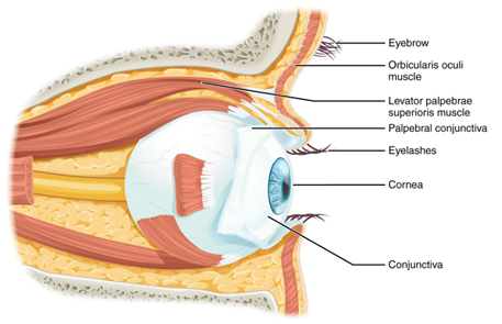
Vision: Eye structure. (Figure by OpenStax is used under a Creative Commons Attribution license.)
Optical Illusions
The brain actively processes visual input, sometimes resulting in optical illusions which are misinterpretations of images caused by surrounding objects, colors, patterns, and preconceptions. Several vision-related activities help illustrate these effects:
- Afterimages: Afterimages occur due to the adaptation and fatigue of photoreceptors in the retina. When you stare at a bright or colored image for an extended period, the corresponding photoreceptors (cones) become desensitized. When you shift your gaze to a blank surface, you perceive a negative afterimage in complementary colors. This phenomenon demonstrates the way our eyes and brain adapt to prolonged stimuli and compensate when the stimuli is removed.
- Benham’s Disk: Benham’s disk is an optical illusion that produces illusory colors from a spinning black-and-white pattern. Even though the disk contains no actual colors, rapid motion activates different cone pathways at slightly different speeds, creating a perception of faint colors. This activity demonstrates how the visual system processes temporal changes in stimuli and how the brain interprets incomplete visual information.
- Blind Spot: The blind spot is an area on the retina where the optic nerve exits the eye, and no photoreceptors are present. In this activity, you will locate your blind spot by focusing on a specific object while another disappears from view as it falls within this region. Normally, we do not notice our blind spot because the brain fills in the missing information based on surrounding visual cues and input from the other eye. This activity highlights how the brain actively reconstructs visual perception to create a seamless experience.
By exploring these visual phenomena, we gain a deeper understanding of how the eyes and brain work together to interpret and process the world around us.
Smell and Taste
Humans are estimated to distinguish over 10,000 different smells (odorants), though describing scents is more challenging compared to describing features of other senses, like color or sound. Despite this vast olfactory capability, only a small patch of specialized cells at the top of the nasal cavity in each nostril is responsible for detecting odors. This regions is known as the olfactory epithelium, contains millions of olfactory neurons equipped with tiny hair-like projections called cilia. These cilia have receptors that bind to specific odor molecules (odorants) in the air. Once an odorant binds to a receptor, the neuron sends a signal via the olfactory nerve to the olfactory bulb, where the information is processed and relayed to the olfactory region of the temporal lobe for interpretation. Over time, continuous exposure to an odor results in decreased sensitivity, a phenomenon known as olfactory fatigue.
To explore how different scents interact, students will smell two different cups. Once cup contains only vinegar, while the other contains a mixture of vinegar and peppermint extract. This activity demonstrates odor masking, where a strong or pleasant smell can suppress the perception of an otherwise dominant odor. You will compare how each sample smells and discuss how the brain processes multiple odor signals at once.
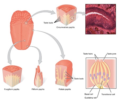
Taste: Chemoreceptors responding to chemicals. (Figure by OpenStax is used under a Creative Commons Attribution license.)
In contrast, the sense of taste is relatively limited, with five primary taste sensations: sweet, sour, salty, bitter, and umami (a savory taste identified in more recent research). Despite having only a handful of taste categories, humans experience a vast range of flavors because much of what we perceive as taste is actually due to olfaction, the smell of food molecules traveling through both the nasal passages and a connection at the back of the mouth to the olfactory epithelium.
Taste perception begins when tastants (dissolved food molecules) interact with specific taste receptors on the tongue. These receptors send signals via sensory nerves to the thalamus, which then relays the information to the cerebral cortex, where the brain interprets the combination of taste and aroma as flavor. Factors such as food texture, prior experiences, and even sound can influence how we perceive taste.
Students will sample five different sport drinks with added food coloring to investigate how visual cues affect taste perception. Each drink will be flavored differently, but the colors may not correspond tot he expected flavors. You will record your guesses and compare them to the actual flavors, highlighting how visual perception influences taste expectations. This activity demonstrates the brain’s reliance on multiple sensory inputs to interpret flavor and how expectations can shape our perception.
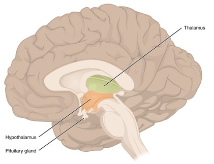
The Brain: Thalamus, hypothalamus, and pituitary gland. (Figure by OpenStax is used under a Creative Commons Attribution license.)
Questions
-
- Describe the functions and components of the following parts of the nervous system:
- The integrating center that receives incoming afferent sensory information and responds by sending out motor signals if changes are needed.
- The system made up of all the afferent, incoming sensory information and efferent, outgoing motor signals to/from neurons outside the CNS.
- Explain the following processes related to sensory perception:
- The process by which stimulus information is transduced (converted) into electrical and chemical signals sent to the brain.
- The process by which the brain “makes sense of” or categorizes the incoming sensory information.
- Describe the function of each type of sensory receptor:
- Photoreceptor
- Chemoreceptor
- Thermoreceptor
- Mechanoreceptor
- Nociceptor
- Explain why visual “tricks” or optical illusions are a result of perception rather than sensation.
- Describe the functions and components of the following parts of the nervous system:
-
- Explain the phenomenon where the sense of smell decreases in sensitivity after continuous exposure to an odor. What is this process called?
Adapted from Human Physiology Lab Manual by Jim Blevins, Melaney Farr, and Arleen Sawitzke, Salt Lake Community College.
