
5 Background: The Cell
Last week we practiced using a compound light microscope and a dissecting microscope and investigated how magnification affects field of view, illumination, depth of field and resolution. This week, our goal will be to compare the cells from different organisms representing the three domains of life. We will focus on the sizes and shapes of cells and on identifying the subcellular structures that we can see under the microscope.
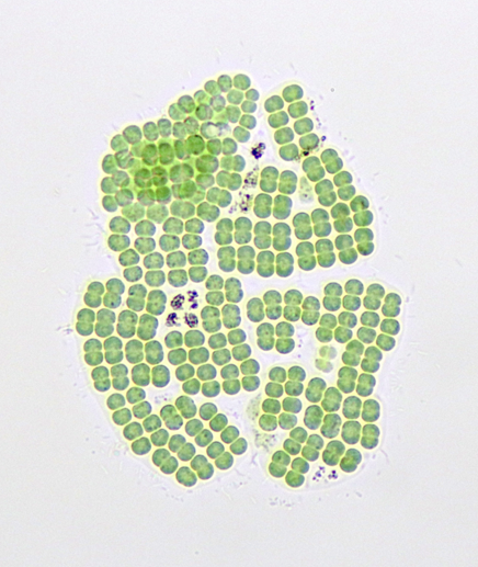
Prokaryotes, unicellular organisms lacking a nucleus, include cyanobacteria (formerly blue-green algae. This name is now considered inaccurate because algae are eukaryotes. Cyanobacteria, like those shown in the figure below, are photoautotrophs—organisms that carry out photosynthesis by using light energy, water, and carbon dioxide from the air and converting to sugars, and providing oxygen to the atmosphere as a waste product. Cyanobacteria contain pigments capable of capturing light energy but do not contain chloroplasts. Cyanobacteria are single-celled organisms, but some can form colonies with differentiated cell types. These cells range from 1–40 micrometers in size. Not all bacteria can carry out photosynthesis there are many species of heterotrophic bacteria living virtually everywhere on Earth. These cells
are much smaller ranging from 0.5–8 micrometers. Eukaryotic cells have many more features and organelles and range in size from 10–500 micrometers
Unlike prokaryotic cells, eukaryotic cells have: 1) a membrane-bound nucleus; 2) numerous membrane-bound organelles such as the endoplasmic reticulum, Golgi apparatus, chloroplasts, mitochondria, and others; and 3) several, rod-shaped chromosomes. Because a membrane surrounds eukaryotic cell’s nucleus, it has a “true nucleus.” The word “organelle” means “little organ,” and, as we already mentioned, organelles have specialized cellular functions, just as your body’s organs have specialized functions.
Animal cells are eukaryotic and possess subcellular components in common with the plant cells. Organelles that plant and animal cells share in common include the nucleus, Golgi apparatus, mitochondria, ribosomes, and the endoplasmic reticulum. These are all participants in protein synthesis. An illustration of an animal cell is shown below. There are some exceptions to these general components. For example, mature red blood cells (RBC) which have ejected their nuclei to have more room for hemoglobin, the protein that carries oxygen around the body. One of the easiest eukaryotic cells to obtain in the lab is the squamous epithelial cell (your cheek cells). These cells are arranged in a flat layer and are easy to remove and observe.
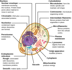
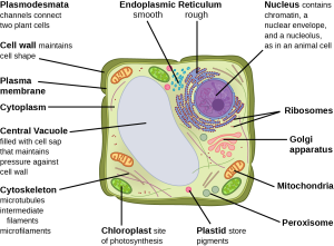
At this point, you know that each eukaryotic cell has a plasma membrane, cytoplasm, a nucleus, ribosomes, mitochondria, peroxisomes, and in some, vacuoles, but there are some striking differences between animal and plant cells. While both animal and plant cells have microtubule organizing centers (MTOCs), animal cells also have centrioles associated with the MTOC: a complex we call the centrosome. Animal cells each have a centrosome and lysosomes; whereas, most plant cells do not. Plant cells have a cell wall, chloroplasts and other specialized plastids, and a large central vacuole; whereas, animal cells do not.
Like the mitochondria, chloroplasts have their own DNA and ribosomes, but chloroplasts have an entirely different function. Chloroplasts are plant cell organelles that carry out photosynthesis. Photosynthesis is the series of reactions that use carbon dioxide, water, and light energy to make glucose and oxygen. This is a major difference between plants and animals. Plants (autotrophs) are able to make their own food, like sugars used in cellular respiration to provide ATP energy generated in the plant mitochondria. Animals (heterotrophs) must ingest their food.
Text and figures in this section adapted from Openstax. Access for free at https://openstax.org/books/biology-2e/pages/1-introduction
